Microstructure and Function (S1)
No description yet. This curated deck is ready for daily study.
Card preview (21)
What is muscle?
any contractile tissue
What are the 5 types of tissue?
• epithelium • connective • blood • muscle • neural
What are the 3 main muscle types?
• Striated • Skeletal • Cardiac • Non-striated: • Smooth
What are other non-striated muscles apart from smooth?
• myoepithelium (in glands, iris of eye) • myofibroblasts (in healing wounds)
How quick is contraction and voluntariness within each of the 3 muscles?
• Skeletal • rapid contraction, subject to fatigue, voluntary • Cardiac • quite rapid, resists fatigue, involuntary • Smooth • slower but powerful, energy-efficient, little fatigue, involuntary (apart from internal bladder sphincter)
Describe the microstructure arrangement of skeletal muscle
• Epimysium (thick outermost CT layer surrounding muscle) • Perimysium (CT layer surrounding fascicles) • Within fascicle, there is a muscle fibre surrounded by endomysium and sarcolemma • Furthest subdivision = myofibrils within the muscle fibre cells
Why are skeletal muscle fibres multinucleated?
Skeletal muscle fibres are formed by thousands of precursor cells in the embryo (myoblasts) fusing together.
What is a myofibril?
Bundles of thick myosin and thin actin filaments
What is a sarcomere?
Contractile unit, from one Z line to the next
What is the Z line?
Where actin filaments meet / separating sarcomeres
What are the A and I bands?
A band is in the middle of a sarcomere, where the myosin and actin filaments overlap. The I band is between the myosin filaments (so just the actin)
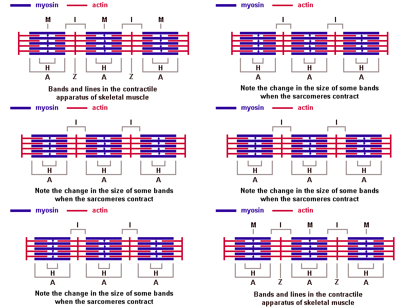
What happens during the sliding filament model?
The sarcomere contracts - the Z lines get closer so the I band gets narrower but the A band stays the same.
How does skeletal muscle know when to contract?
There is neural stimulation via motor end-plates, special membrane systems carry the signal to all parts of this very large cell.
What is Duchenne muscular dystrophy?
Inherited genetic condition in whcih the muscle fibres are smaller and there is more connective tissue.
What are the main differences between cardiac and skeletal muscle fibres?
Cardiac has much smaller fibres, joined end-to-end by specialised junctions called intercalated disks, fibres also only have 1 or 2 nuclei.
How does cardiac muscle resist fatigue?
The numerous mitochondria allow continuous supply of energy
How does cardiac muscle resist high blood pressure?
It has branching fibres which provide extra strength
Describe the myogenic stimulus of cardiac muscle?
AP starts from pacemaker in R. Atrium and carries wave of contraction across heart, assisted by ion diffusion through gap junctions in intercalated disks.
Where is smooth muscle found?
Generally internal organs (except heart), eg. digestive tract, uterus, lungs, bladder, glands, blood vessels etc
What is the shape and structure of smooth muscle fibres like?
smooth, sausage spindle shaped, with one nucleus
What does the contractile structure of smooth muscles look like?
No myofibrils/striations/complex membrane systems, but there are caveolae and ER. Also some actin and very few myosin filaments.