2. Cross sections
No description yet. This curated deck is ready for daily study.
Card preview (4)
eye
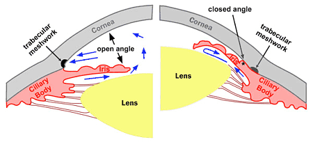
\the three layers of the eyeball consist of the outer fibrous sclera, the middle vascular choroid and the inner layer of the retina. The sclera is continuous posteriorly with the dural cuff that surrounds the optic nerve, and is continuous anteriorly with the cornea. Contents of the globe: the posterior part contains vitreous humour, which is a colourless transparent gel and which constitutes about 80% of the total contents. Anteriorly, the vitreous body is bounded by the capsule of the lens and its suspensory ligaments. These ligaments extend to the ciliary body, which is a direct anterior continuation of the choroid and comprises the muscle involved in accommodation of the lens. The ciliary body also secretes aqueous humour into the anterior chamber, where it circulates before draining via spaces in the trabecular meshwork through the canal of Schlemm. The anterior chamber of the eye contains the iris, which itself is a forward continuation of the choroid via the ciliary body.
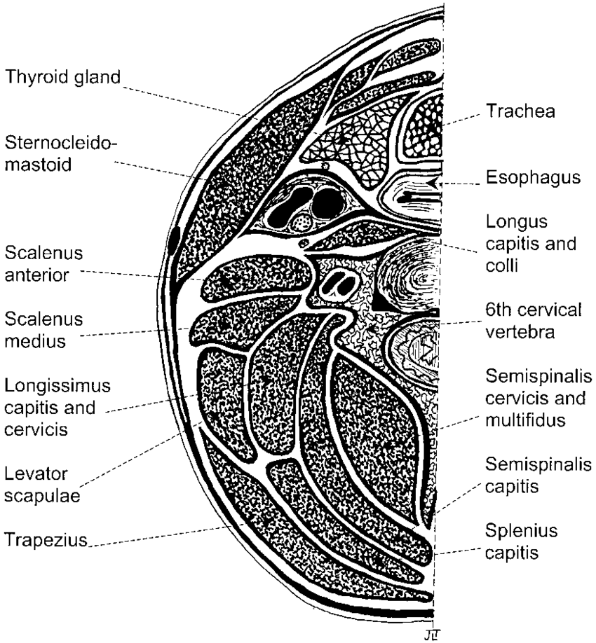
The Neck at the Level of C6
C6 is the level of the cricoid cartilage whose lower border marks the beginning of the trachea. Immediately posterior is the oesophagus, which is separated from the body of the sixth cervical vertebra only by the pretracheal fascia Immediately anterior is the isthmus of the thyroid. Posterolaterally is the carotid sheath, which encloses the common carotid artery, the internal jugular vein and the vagus nerve. Behind the sheath lies part of the sympathetic chain. Immediately lateral to the vertebral body are the vertebral artery and vein, beyond which are the scalene muscles. Between these lie the trunks of the brachial plexus.
The Lumbar Region at the Level of L3
Immediately anterior to the vertebral body are the aorta (on the left) and the inferior vena cava (on the right). On the lateral surface on each side lies the sympathetic trunk. Immediately lateral to the vertebral body at this level lies the psoas major muscle. Further lateral are the lower poles of the kidney. Posterior to the vertebral body is the vertebral canal, which, at the level of L3, contains the theca (comprising the dura and arachnoid maters), within which are CSF and the cauda equina. The theca is surrounded by the epidural space. This is minimal anteriorly because the dura is closely apposed to the vertebral body. The vertebral arch is completed by the pedicles, transverse processes, laminae and spinous processes.
The Lower Thoracic Region at the Level of T10
Remember that you are effectively looking up at the image, and so the left side of the patient is on the right as you view it. The structures are as identified on the CT image reproduced in Figure 2.15 and show the liver, stomach, spleen and blood vessels.