2.4 Ocular Anatomy
No description yet. This curated deck is ready for daily study.
Card preview (22)
The Anatomy of the Orbit
The bony orbit has been described variously as a pyramid whose apex is directed inwards and upwards, as a cone and as a pear whose stem points towards the optic canal. Its roof consists of the orbital plate of the frontal bone, with the anterior cranial fossa above, while its floor is formed by the zygoma and the maxilla, with the maxillary sinus beneath. Its medial wall is formed by parts of the maxilla, lacrimal bone, ethmoid and sphenoid, and beyond it lie the ethmoid air cells and the nasal cavity. The zygoma and the greater wing of the sphenoid make up its lateral wall.
Contents of Orbit
The bony orbit contains the globe, together with the muscles, nerves and blood vessels that subserve the normal functions of the eye.
Normal length + layers
The normal globe has an axial length of around 24 mm (as measured in the anteroposterior diameter). An eye longer than 26 mm is usually myopic. Its outer layer comprises sclera and cornea; the middle vascular layer contains the choroid, the ciliary body and the iris; and the innermost layer comprises neural tissue in the form of the retina.
The movements of the globe
The movements of the globe are controlled by the six extraocular striated muscles. The four recti (lateral, medial, superior and inferior) originate from the annulus of Zinn, the tendinous ring which encircles the optic foramen, and insert beyond the equator of the globe. The lateral and medial recti have two heads. The superior oblique muscle originates above and medial to the annulus, curves round the trochlea (which acts like a pulley), before inserting behind the equator and beneath the superior rectus. The inferior oblique originates from the lacrimal bone and inserts posterolaterally on the globe, having passed beneath the inferior rectus muscle.
Motor innervation:
the lateral rectus is supplied by the sixth cranial nerve, the abducens (VI), and the superior oblique is supplied by the fourth, the trochlear (IV). SO4\LR6 The remaining muscles are supplied by the third cranial nerve, the oculomotor (III). (This also supplies levator palpebrae superioris, which elevates the eyelid).
Autonomic innervation
Autonomic innervation: sympathetic innervation is by the long and short ciliary nerves via the superior cervical ganglion. Nerve impulses dilate the pupil via the dilators of the iris. Parasympathetic innervation is by the short postganglionic ciliary nerves via the ciliary ganglion. The pre-ganglionic supply comes from the oculomotor nerve, and its impulses constrict the pupil.
Sensory supply:
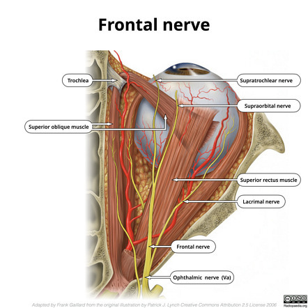
this is derived mainly from the ophthalmic branch of the fifth cranial nerve, the trigeminal (V), although branches of the maxillary division make some contribution to lateral structures and to the nasolacrimal apparatus. There are a large number of sensory nerves for such an anatomically confined area. The examiner is unlikely to dwell on these in any detail but, in summary, the innervation that may have relevance for ocular surgery can be outlined as follows. The ophthalmic division V1 branches into the frontal nerve, which then divide:
Frontal N Branches
• supratrochlear nerves (medial upper conjunctiva), • the supraorbital nerve (upper conjunctiva) • the long ciliary nerve (cornea, iris and ciliary muscle).
V1
V1 also forms the nasociliary nerve, which in turn branches into the infratrochlear nerve (inner canthus and lacrimal sac) and the long sensory root to the ciliary ganglion (thence to the cornea and iris). The lacrimal branch of V1 supplies the rest of the conjunctiva.
Foramina Optic canal
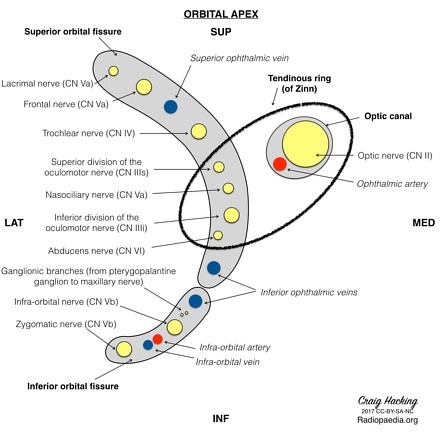
Foramina: the orbit contains nine fissures and foramina, of which three are particularly important: 1. the optic foramen (canal), 2. the superior orbital fissure 3. inferior orbital fissures.
Optic canal
. The optic nerve and ophthalmic artery traverse the optic foramen.
Superior orbital fissure
Through this fissure run the oculomotor, trochlear and abducens nerves to the extraocular muscles, together with the frontal, nasociliary and lacrimal nerves and the superior and inferior ophthalmic veins. The oculomotor, abducens and nasociliary nerves traverse the lower part of the fissure and enter the muscular cone between the two heads of the lateral rectus. The trochlear, frontal and lacrimal nerves remain outside the cone.
• Inferior orbital fissure
Through the inferior fissure run the zygomatic and infraorbital nerves (branches of V2), the infraorbital artery and the inferior ophthalmic vein.
Ophthalmic Reflexes
• Corneal reflex • Pupillary Reflex - miosis - mydriasis • Abnormal Sx
Corneal reflex:
This is the normal blink reflex but is used as part of brain stem death testing. Stimulation should provoke both a direct and a consensual reflex. The afferent pathway from the nasociliary branch of the ophthalmic division of the trigeminal nerve (V1) leads to the trigeminal sensory nucleus in the medulla oblongata. Interneurons connect to the facial motor nucleus which mediates the motor response (contraction of the orbicularis oculi muscles) via the temporal and zygomatic branches of the facial (seventh cranial) nerve.
Pupillary reflexes: constriction (miosis).
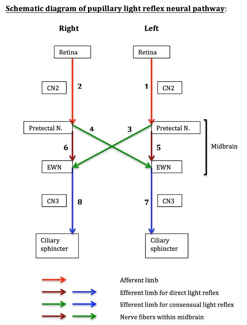
These are essentially photopupillary responses to the intensity of ambient light. In health, this reflex is also consensual; thus stimulation of one pupil elicits the same response in the other. The sensory afferent apparatus consists of photosensitive cells in the retina, the optic nerve and the pretectal nucleus in the midbrain. Efferents from the pretectal nucleus pass to the ipsilateral and contralateral Edinger-Westphal nuclei (also in the midbrain). From these nuclei project preganglionic parasympathetic fibres which run with the oculomotor (third cranial) nerve to synapse with post-ganglionic neurons in the ciliary ganglion. The short ciliary nerves (around 6–10 in number) project from this ganglion to innervate the sphincter pupillae of the iris. Stimulation of this muscle results in pupillary constriction (miosis).
Abnormal pupillary signs.
These are many in number and include eponymous rarities such as the • Holmes-Adie syndrome (sluggish pupillary reflexes secondary to denervation in postganglionic parasympathetic nerves) and the • Argyll-Robertson pupil of tertiary syphilis (no constriction in response to light, but constriction in response to accommodation).
if a patient complains of unilateral eye pain with a dilated unreactive pupil
if a patient complains of unilateral eye pain with a dilated unreactive pupil, and a contralateral pupil that constricts directly and consensually (with no other signs), then it is possible to deduce that the problem involves motor innervation at the level of the ipsilateral ciliary ganglion or short ciliary nerves. The oculomotor nucleus and nerve are not involved because normal eye movements are not affected.
Local Anaesthesia for Eye Surgery
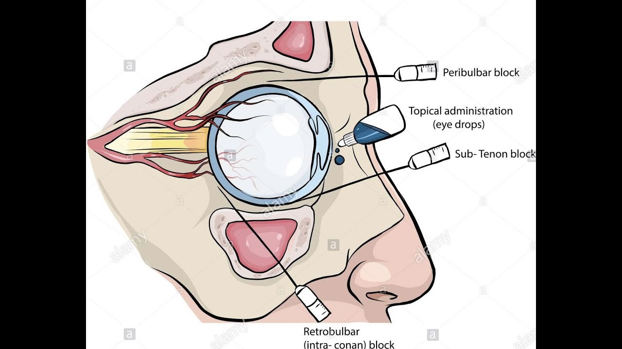
You may be asked about methods of anaesthetizing the eye for intraocular surgery. Although retrobulbar and peribulbar blocks are being supplanted by sub-Tenon’s block and by topical local anaesthesia, they allow some discussion of the anatomy. You will only have to discuss one or two of these methods, usually the one(s) with which you are familiar, and so there is more detail in what follows than you will need.
Topical:
the anterior structures can be anaesthetized using topical amethocaine 0.5% or 1.0%, oxybuprocaine 0.4% and proxymetacaine 0.5%. Topical anaesthesia is simple and (mostly) safe and effective, although the lack of akinesia of the eye and eyelids means that the surgeon has to control eye movement via the intraocular instruments. Anaesthesia can be supplemented by the addition of lignocaine to the irrigation fluid, or by further instillation of drops. These can cause oedema of the cornea, and excessive doses may exacerbate the problem.
Sub-Tenon’s block
: the popularity of this technique has increased because it is viewed as safer than the sharp needle approaches. It is, however, more invasive, in that a modest amount of surgical dissection is necessary. After topical anaesthesia to the conjunctiva the patient is asked to look upwards and outwards (in the direction of the operator). This improves access to the inferonasal quadrant where the injection is made, as posteriorly as possible fold of conjunctiva is drawn upwards with forceps. A small nick at the base of this fold with surgical scissors opens the sub-Tenon’s fascia. blunt cannula is then inserted gently into this space and guided backwards following the contour of the globe. Injection of 4–5 ml of local anaesthetic solution will provide analgesia and adequate akinesia. This injection requires pressure to distend the space and some patients find this very uncomfortable. The globe can in theory be perforated, and central spread of local anaesthetic has been described, but these complications are sufficiently rare for sub-Tenon’s block to be considered suitable for administration by trained, but non-medical, practitioners.
Peribulbar block:
this has been cited as a safe and effective alternative to retrobulbar block, but it too is not without its problems. Larger volumes of local anaesthetic are required (8–10 ml), which increases the intraorbital pressure and causes periorbital chemosis. The onset of block is also considerably slower and the failure rate higher. The risk of scleral perforation is not removed because the technique requires one inferotemporal and one superonasal injection, both of which are directed beyond the equator of the globe. (Some include a third injection, made at the extreme medial side of the palpebral fissure).