Proteins: Structure & Function
No description yet. This curated deck is ready for daily study.
Card preview (18)
What are proteins and how are they formed?
Each protein has a unique and specific amino acid sequence that is genetically determined by the order of nucleotide bases in the DNA (the genetic code). Proteins are formed from amino acids joined together by peptide bonds or amide bonds.
How are peptide bonds formed?
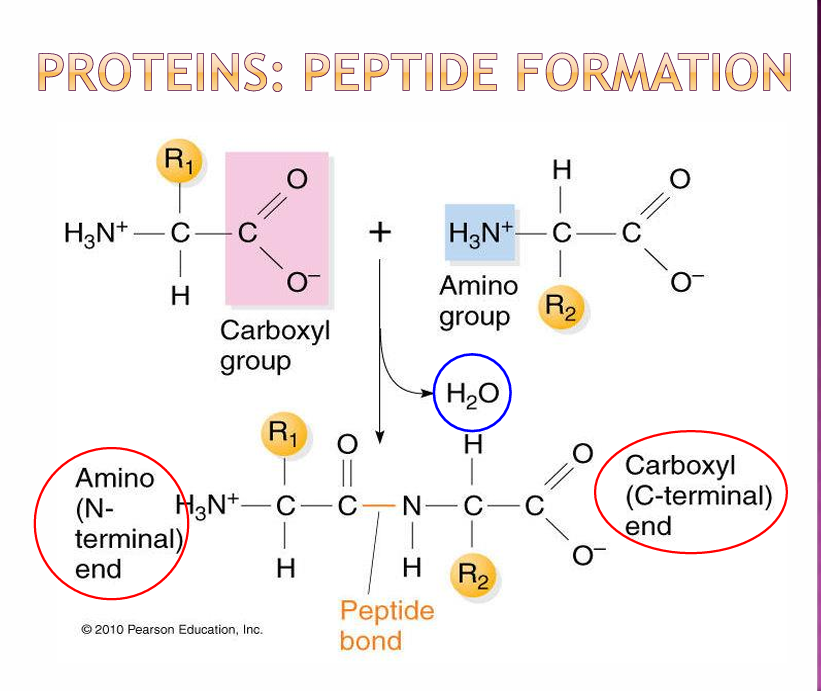
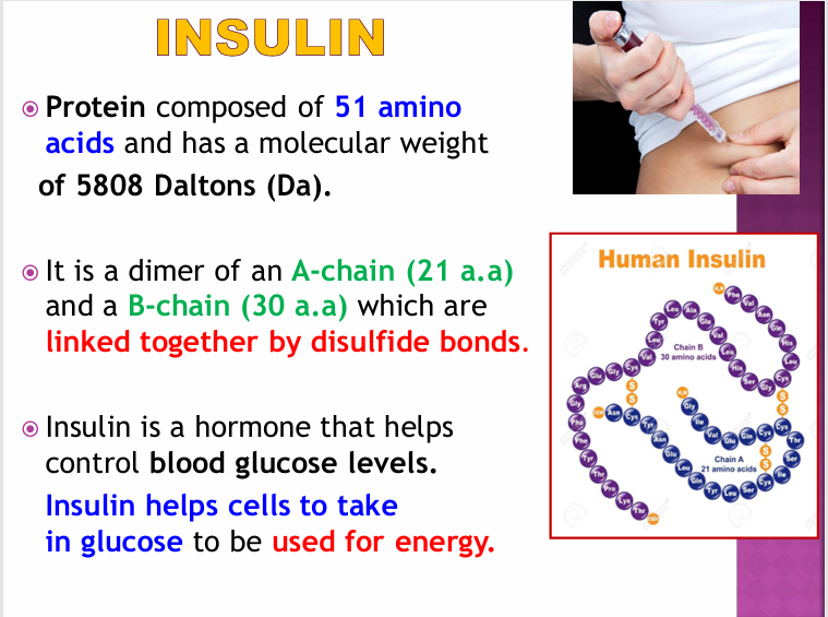
Peptide bonds are formed by a condensation reaction (remove H2O) of a carboxylic group of an amino acid an amino group of another amino acid. Dipeptides (2), tripeptides (3) ,oligopeptides (5-10) amino acids; polypeptides (<50 amino acids). Proteins have >50 amino acids. Insulin is an exception as it is a polypeptide with 51 amino acids.
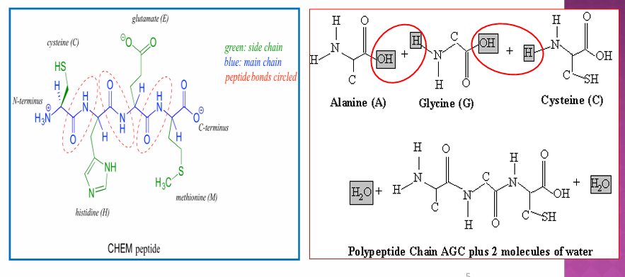
Insulin
What are thr four levels of protein organisation?
• Primary Structure • Secondary Structure • Tertiary Structure • Quarternary Structure
Please explain Primary and Secondary structure
• Primary Structure - linear amino acid sequence of polypeptide chain; determined by genetic code and held by peptide bonds. • Secondary Structure - interactions between adjacent amino acids. The formation of a regular pattern of twists or kinks of the polypeptide chain. Held together by hydrogen bonds. Eg. Alpha helix - stabilised by hydrogen bonding between carbonyl atom and amide hydrogen. It has a spiral structure which pulls the polypeptide chain into a helical structure like a curled ribbon. Each turn of the helix contains 3.6 amino acids. Beta pleated sheets - 2/more segments of the polypeptide chain line up to each other to form a sheet-like structure held by hydrogen bonds. The strands of the B-pleated sheet may run parallel or anti-parallel to each other.
Explain Tertiary Structure?
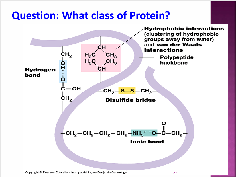
Tertiary Structure - 3D folding of polypeptide. The linear sequence of the amino acid is folded into a compact globular structure. The folding and final shape of the polypeptide chain is determined and stabilised by a variety of interactions between R groups. These interactions include hydrogen bonds, electrostatic interactions, hydrophobic interactions (the folded protein is soluble in water or aqueous solutions) and covalent bonds and di-sulphide bonds. Eg. Cysteine has an SH group as part of its R group and forms di-sulphide bonds with itself.
Explain Quarternary Structure
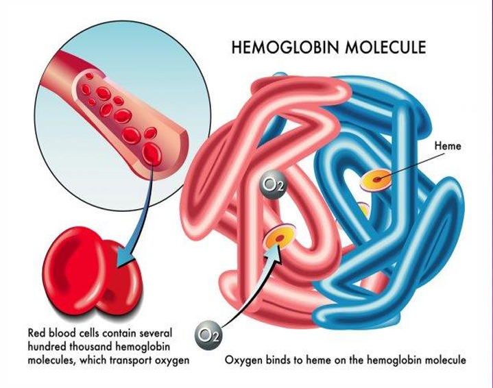
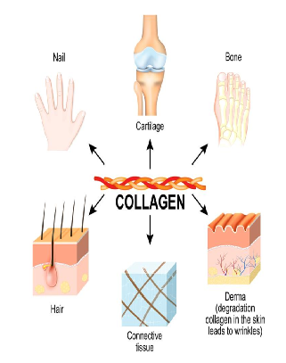
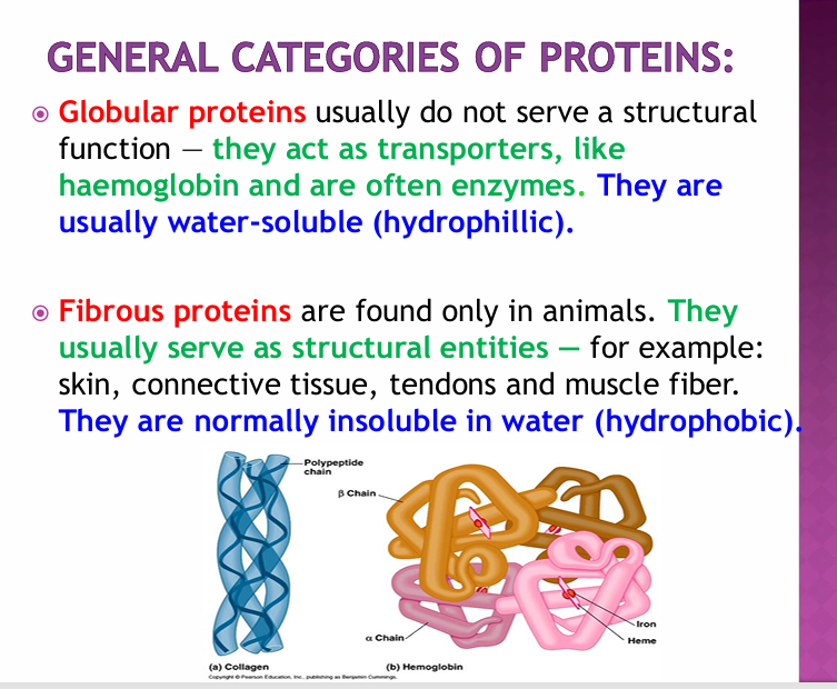
Quarternary Structure - arrangements of multipe polypeptides. They contain 2/more tertiary subunits and form either globular or fibrous proteins. Eg. Haemoglobin: globular protein that contains 2 alpha chains and 2 beta chains. The haeme group in each subunit transports the oxygen in the blood. Collagen is a fibrous protein made of 3 polypeptides supercoiled like rope which provides strucutral strength for connective tissue. Quaternary is dependent on the tertiary structure of the individual polypeptides and so is influenced by these bonds.
Globular vs Fibrous proteins
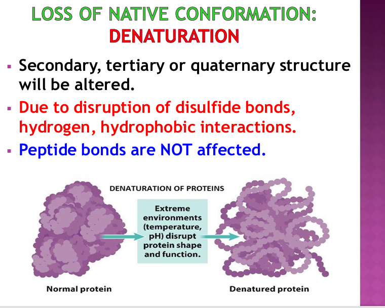
Denaturation
Protein Functions:
• Enzymes: proteins that increase the rate of chemical reactions in the body. • Structural: Skin and bone contain collagen, a fibrous protein. • Regulatory: involved in the creation of some hormones which help to control body functions that involve the interaction of several organs and help to regulate cell growth and body functions. • Transport: Hb • Muscle fibres • Help store other substances: Iron is stored in the liver in a complex with the protein ferritin. • Antibodies: proteins that help to rid the body of foreign invaders eg. Bacteria, viruses etc and thus help to prevent infections, illnesses and diseases. Year 2020: COVID-19 Pandemic. Total length of SARS-CoV-2 Spike protein is 1273 aa’s
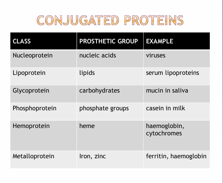
What are conjugated proteins?
Conjugated proteins are complex molecules consisting of amino acids covalently bonded or weakly interacted with non-protein components known as prosthetic groups.
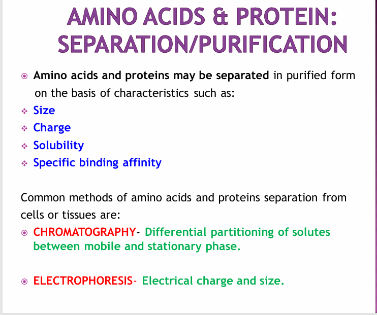
Amino Acid Separation and Purification
Types of Chromatography
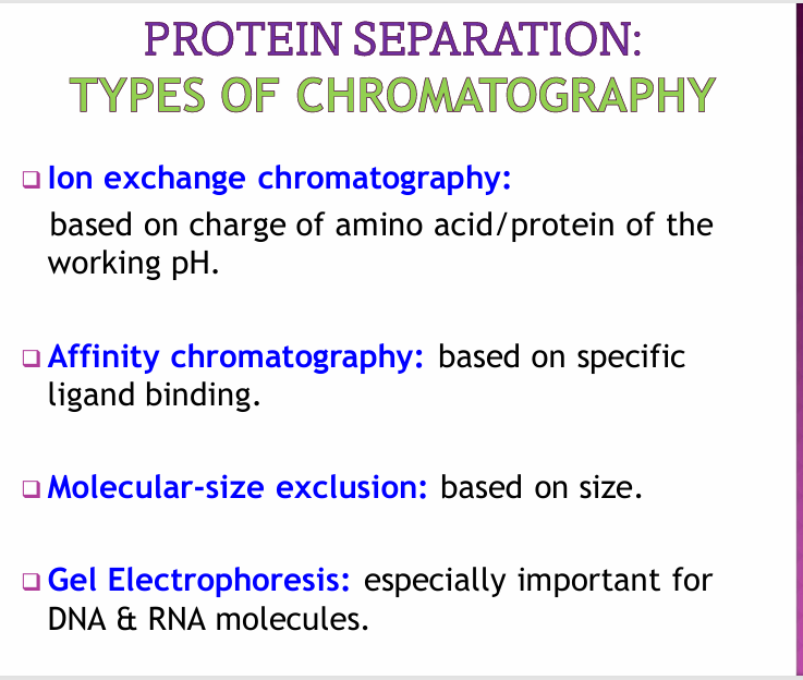
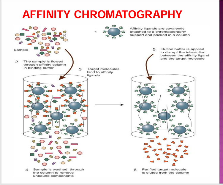
Ion-exchange and affinity chromatography
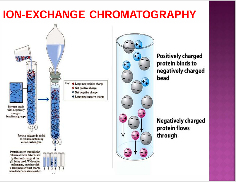
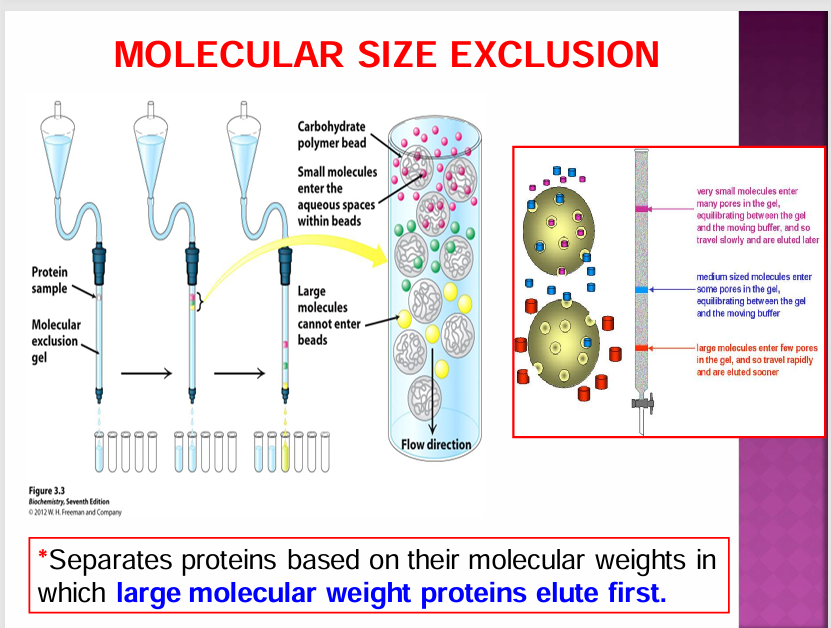
Molecular (Size) Chromatography
Gel Electrophoresis
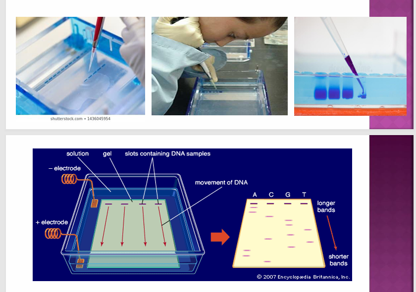
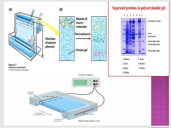
Separates fragments of DNA, RNA and even proteins based on their molecular weight. Process requires a well of gel where an electric field is introduced which induces movement of the target molecules based on their charge and also size because of the molecular sieving effect of the gel. DNA and RNA are negatively charged molecules so they will be pulled towards the positively charged end of the gel. Different types of gels which can be used depending on use and application: • Agarose gel- very large proteins and nucleic acids • Polyacrylamide gel- small proteins and nucleic acids After the electrophoresis is complete, the molecules in the gel can be stained to make them visible. Ethidium bromide, silver or coomassie blue dye may be used for the process. Other methods may be also used to visualize the separation of the mixture’s components on the gel: If the analyte molecules fluoresce under ultraviolet light, a photograph can be taken of the gel under ultraviolet lighting conditions.
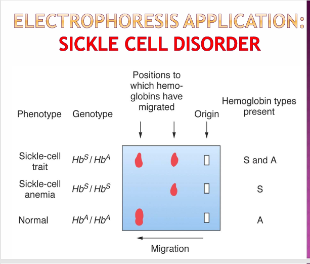
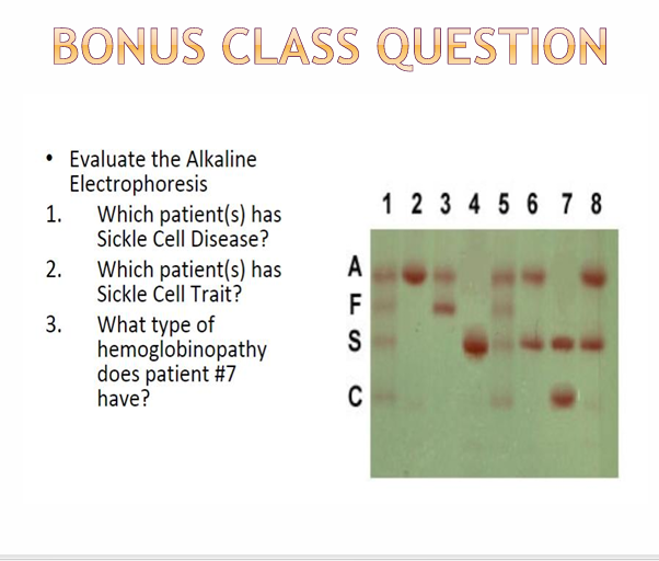
Sickle Cell
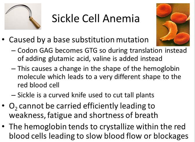
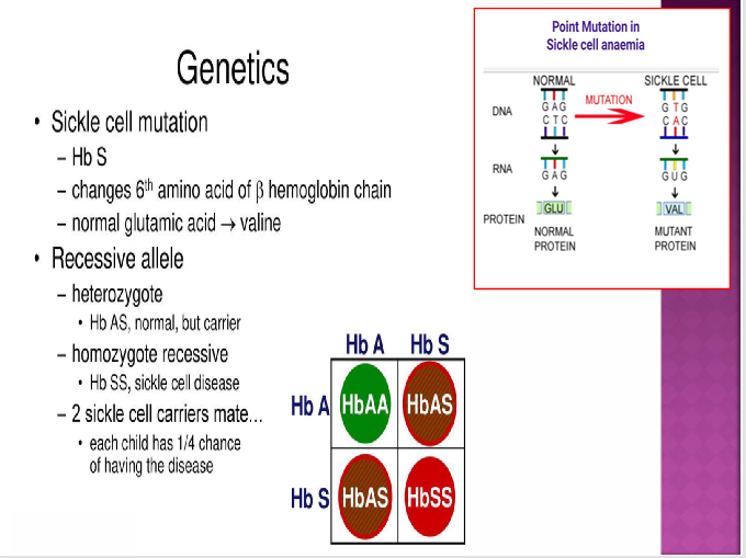
A change in just one amino acid can change the structure and function of a protein. For example, Sickle cell anaemia is a disease that results from an altered structure of the protein haemoglobin (in the beta globin chain). At 6th position the hydrophilic glutamic acid gets replaced with the hydrophobic valine. This is the result of a point mutation- single base pair change at the DNA level.
Electrophoresis and Sickle Cell