Smooth Muscle
No description yet. This curated deck is ready for daily study.
Card preview (14)
Where is smooth muscle?
Hollow organs + tubes Helps regulate flow in: • vascular system • airways • GI tract • urogenital tract • eye
Functional roles of smooth muscle
Regulate flow by varying tube diameter - blood vessels during exercise Control flow by occluding tube - sphincters Walls of storage organ - expand + expel Movement of large bulk - peristalsis (oesophagus)
Describe the structural organisation of smooth muscle
Supported by and contains connective tissue SM does not act on structures like bone (has no tendons) Small cells (do not extend full length of muscle) Group of cells arranged in sheets Single sheet e.g. arterioles + airways • circularly oriented • maintains vessel diameter • controls blood flow Multiple sheets e.g. ileum • 2 sheets perpendicular to each other • longitudinal + circular layers • vary diameter + length: peristalsis
Describe smooth muscle cell structure
Small spindle-shaped cells • Uni-nucleate • No striations, not banded i.e. “smooth” • No “z-bands” but “dense bodies” Actin & myosin filaments present • actin filaments anchored to “dense bodies” Intracellular cytoskeleton harnesses pull • Contracts inward, bulges • Intercellular connections harness pull between cells
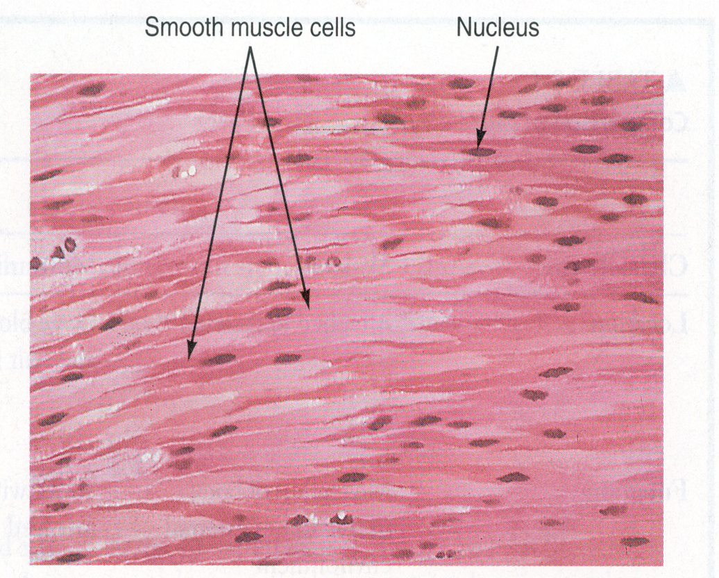
What are Gap Junctions?
Join smooth muscle cells to one another Non-selective channels allow intracellular communication Signalling propagate between cells Fibres act in unison: synchronised contraction of uterus during labour
What is needed to start a SM contraction?
Requires Ca2+ from either: • outside cell across plasma membrane • using voltage gated Ca2+ channels • Ca2+ mobilised from intracellular stores • through activation of 2nd messenger Increasing Ca2+ increases the level of tone by increasing the contraction of the SM cells Varying the level of intracellular Ca2+ enables SM cells to vary level of contraction
Example of structure made up of multiple sheets of smooth muscle
Ileum (final part of SI)
Example of structure made up of a single sheet of smooth muscle
Arterioles + airways
Describe the contractile mechanisms of smooth muscle
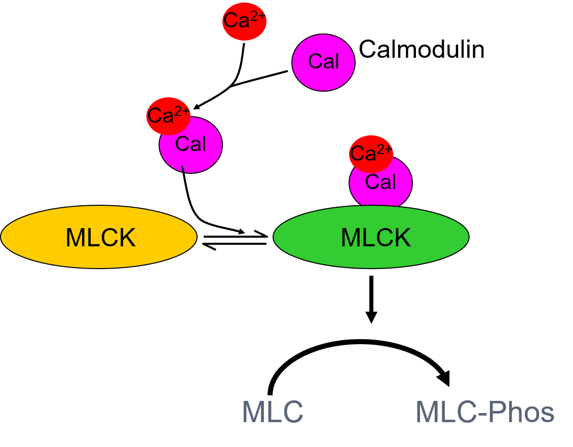
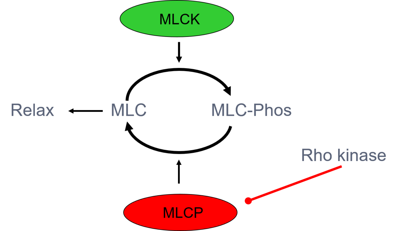
Contraction by sliding filament theory (Interaction of actin w/ myosin to form X-bridges) Ca2+ binds to calmodulin (biochemical effect) Ca2+-calmodulin activates myosin light chain kinase (MLCK) MLCK phosphorylates myosin light chain (MLC) MLC must first be phosphorylated for actin to bind This causes a contraction
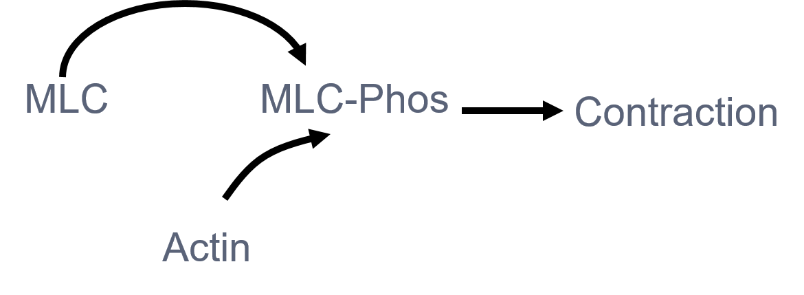
How can MLC bind to Actin
MLC must be phosphorylated to MLC-Phos to be able to bind to Actin to cause a contraction
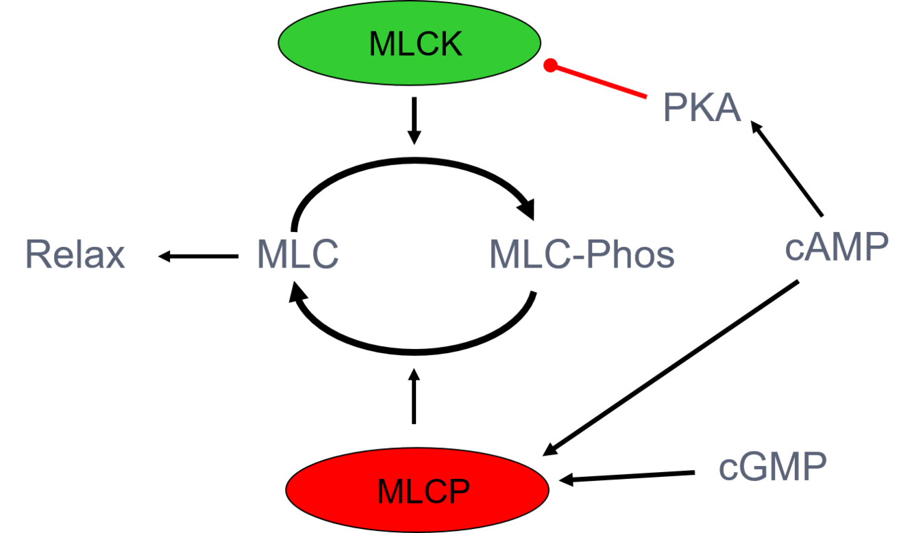
Relaxation of smooth muscle
Ca2+ in cytosol decreases Ca2+ unbinds from calmodulin MLCK decreases MLCP (myosin phosphatase) removes phosphate group from myosin light chains which decreases myosin ATPase (MLCP from cGMP or cAMP) Less myosin ATPase causes decreased muscle tension
How does Rho-Kinase play a part in smooth muscle contraction?
RhoA activates Rho kinase, which phosphorylates MLCP, thereby inhibiting MLCP activity and increasing contraction and vascular tone.
What receptors increase cAMP in smooth muscle?
Beta-adrenoceptors Increasing cAMP leads to relaxation (airways) No relaxation of vascular smooth muscle (nitrates used in angina)
What are the other ways which smooth muscle tone can be regulated?
By contractile + relaxatory agents released from neurones, endothelium + blood-borne