2.11 Cervical Plexus
暂无描述。系统推荐的高质量记忆内容,适合每天坚持背诵学习。
卡片预览 (13 张)
Relevance
The question of which of general or local anaesthesia for carotid endarterectomy is the better option was not informed definitively by the GALA trial, not least because lack of funding meant that this was discontinued before the planned 5,000 patients had been recruited and because there was considerable criticism about aspects of its methodology. Clinical practice changed over the years during which patients were recruited, and neither the general anaesthetic nor the regional anaesthetic techniques were standardized. Nonetheless, in conjunction with other evidence it seems clear that in respect of major perioperative outcomes, there is no difference between regional and general anaesthesia, with mortality and stroke rates of around 5% in both groups.
Nerve supply to neck
nerves which supply the lateral aspect of the neck all derive from the ventral rami of the second, third and fourth cervical spinal nerves (C2, 3, 4). The first cervical nerve has no sensory distribution to skin.
Superficial cervical plexus anatomy
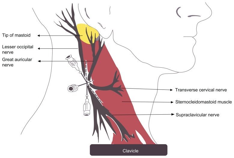
The cutaneous supply to the anterolateral aspect of the neck is via the anterior primary rami of C2, C3 and C4. These nerves emerge from the posterior border of the sternocleidomastoid muscle midway between the mastoid and the sternum. The accessory nerve is immediately superior at this point. The lesser occipital nerve (the first branch) supplies the skin of the upper and posterior ear, the greater auricular nerve (the second branch) supplies the lower third of the ear and the skin over the angle of the mandible, the anterior cutaneous nerve (the third branch) supplies the skin from the chin down to the suprasternal notch and the supraclavicular nerves (the fourth branch) supply the skin over the lower neck, clavicle and upper chest
Superficial cervical plexus block
Superficial cervical plexus block: all these nerves can be blocked at the midpoint of the sternocleidomastoid by infiltrating up to 20 ml of local anaesthetic solution between the skin and the muscle. The external jugular vein crosses the muscle at this point and can be a useful landmark.
Deep cervical plexus anatomy:
Deep cervical plexus anatomy: The ventral ramus of the second nerve emerges from between the vertebral arches of the atlas and axis and runs forwards between their transverse processes to exit between longus capitis and levator scapulae. The ventral ramus of the third nerve exits the intervertebral foramen lying in a sulcus in the transverse process, emerging between the longus capitis and scalenus medius muscles. The ventral rami of the fourth and remaining cervical nerves appear between the scalenus anterior and the scalenus medius.
Deep cervical plexus block:
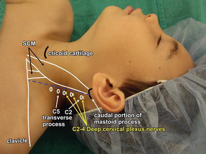
Deep cervical plexus block in effect is a paravertebral block of C2, C3 and C4. Needles are inserted at each of the three levels, using as landmarks a line between the mastoid process and the prominent tubercle of the sixth cervical vertebra (which is palpable as Chassaignac’s tubercle at the level of the cricoid cartilage). The C2 transverse process is approximately one finger’s breadth below the mastoid process along this line with C3 and C4 following at similar intervals caudad. After encountering the transverse process, 5–8 ml of local anaesthetic can be injected with due precautions. Because there is little resistance to the spread of solutions through the paravertebral space in the cervical region, adequate anaesthesia can also be obtained using a single needle technique and a larger volume (15–20 ml) at a single level, usually C3.
Indications for cervical plexus blockade:
these include anaesthesia for carotid surgery under local anaesthesia, clavicular surgery (typically open reduction and internal fixation following trauma) and thyroid surgery.
Advantages of CEA under local anaesthesia
normal cerebration depends on adequate cerebral perfusion, and in the awake patient it is usually obvious whether this is being preserved. In effect the patient acts as their own cerebral function monitor, and signs of cerebral ischaemia are an indication for surgical shunt insertion. Local anaesthesia does not interfere with cerebral autoregulation, and signs of cerebral ischaemia are an indication for surgical shunt insertion. Local anaesthesia does not interfere with cerebral autoregulation, and the requirement for vasoactive drugs is less. Proponents of the technique claimed lower morbidity and mortality rates, but there is no evidence to support that view.
Disadvantages of CEA under local anaesthesia
Disadvantages of CEA under local anaesthesia: cerebral oxygen consumption does not fall (the cerebral metabolic rate for oxygen, CMRO2, decreases under general anaesthesia), and a higher pulse and blood pressure during surgery results in higher myocardial oxygen demand than would otherwise be the case. It does also mean, however, that cerebral perfusion pressure is higher. Cooperation can on occasion be a problem; immobility during extended surgery may be very uncomfortable for the patient and, should their cerebration be obtunded by ischaemia, they may become restless and agitated. The nerve blocks may sometimes prove inadequate as surgery proceeds, but local supplementation by the surgeon can circumvent this problem.
Advantages of CEA under general anaesthesia:
general anaesthesia allows more control, can be extended indefinitely if necessary and during long procedures is more comfortable for the patient. At concentrations up to 1.0 MAC, sevoflurane decreases cerebral blood flow and CMRO2. Experimental evidence suggests that general anaesthetic agents may confer a degree of neuroprotection, but the data are not robust enough to mandate their use.
Disadvantages of CEA under general anaesthesia:
it is clearly more difficult to assess cerebral oxygenation, and, although low concentrations of volatile agents do reduce CMRO2, they may still impair dynamic cerebral autoregulation at MAC levels below 1.0. Monitors of cerebral oxygenation include near-infrared spectroscopy (NIRS), electroencephalography (EEG), somatosensory evoked potentials (SSEPs) and transcranial Doppler. There are in addition the generic complications of general anaesthesia (in which the examiner will have little interest) and those of anaesthesia for head and neck surgery, such as restricted access to the airway.
Complications:
Complications: superficial cervical plexus block risks mainly what can be described as generic complications of local anaesthesia, namely intravascular injection and systemic toxicity. The complications of deep cervical block are much the same as those associated with interscalene block, which is not surprising given the anatomical similarities, and include injection into the vertebral artery, extension of the block either extradurally or intrathecally, phrenic nerve block and cervical sympathetic block, which will manifest as Horner’s syndrome (miosis, ptosis, anhidrosis and enophthalmos). The recurrent laryngeal nerve may also be affected with resultant hoarseness
The GALA trial:
this multicentre trial was conducted over around seven years between 2001 and 2007 and recruited 3.500 of the planned 5,000 patients, who were randomized either to general or regional anaesthesia for carotid endarterectomy. Thereafter, anaesthetists and surgeons were free to follow their routine practice. Primary outcomes were death, stroke or myocardial infarction within 30 days of surgery; secondary outcomes added death at 1 year, length of stay and quality of life. There were essentially no differences between the groups. Criticisms of the trial included the fact that surgical and anaesthetic techniques were very variable; an obvious example being the use or otherwise of shunts, and that both may have changed during the relatively long period during which patients were recruited (GALA Trial [Lancet 2008, 372: 2132–42]).