Unit 1: How we move
No description yet. This curated deck is ready for daily study.
Card preview (24)
1.1 Skeletal system Difference between axial and appendicular skeleton
Axial: structure: middle of our body (medial) ex. ribs, skull, vertebral colum, sternum Function: protection of important structures, support posture, location for muscles to attach Appendicular: structure: all other bones (more lateral) ex. shoulder girdle, pelvic girdle, foot bones function: movement, locations for muscles
1.1 Skeletal system State the different categories of bones four main types of bones
• long bones: * longer than wide * for movement * example: humerus, femur • Flat bones: * curved surfaces * protection and muscle attachment * example: scapula, pelvis, sternum • irregular bones: * specialized shapes and functions * example: vertabrae, sacrum, coccyx • short bones: * cubed shaped * articulate (Connext/move) with +1 other bones * example: tarsals and carpals
1.1 Skeletal system How do bones connect to each other (5)
• joints * a point at which two or more bones articulate * joint or jucture between bones or cartilages in the skeleton * movable joints between rigid parts of an animal • connective tissue: * hold parts of the body together • tendons * attaches muscle to bones (Bottom - bone tendon muscle) * allow for the force generated by the muscle to move the skeleton • ligaments * attaches bone to bone (BLOB - bone ligament bone) * stabilize joints so that the bones stay in the proper location * will be set up in a way that the bones cannot move in a way other than the intended joint movement direction • cartilage * strength and flexibility * found in many joints covering bone * allows for easier movement of the joint as it is smooth * provides some movement (Ears, nose, ribs)
1.1 Skeletal system different types of joints in relation to movement permitted(3)
• fibrous joints * thin fibrous material between the edges of bones * continuous with the surface layer of the bone * NO movement allowed • cartilaginous joints * bones are seperated by a fibrocartilage disc or by a thick layer of cartilage * limited movement in these joints • synovial joints * most common * largest range of motion
1.1 Skeletal System types of synovial joints
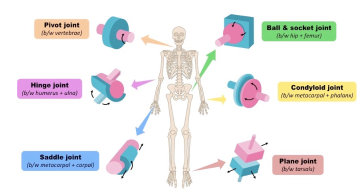
• hinge - permits motion in only one plane (b/w humerus + ulna) • ball and socket - multiple directions of movement (b/w hip + femur) • condyloid - Movement in two directions formed by concave shape fitting into convex shape (b/w metocarpal + phalanx) • pivot - Allows for rotation around the length of a bone, and only allows for rotation. (b/w vertabrae) • gliding - allows one bone to slide over another (b/w tarsals) • saddle - allows grasping and rotation (b/w metacarpals + carpal)
1.1 Skeletal system Structure of synovial joints (7)
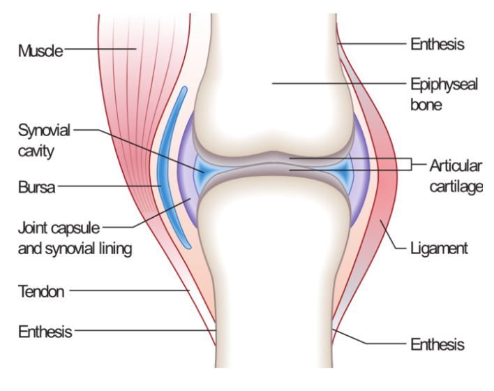
• Articular cartilage → reduce friction, absorb shock • Synovial membrane → produce synovial fluid • Synovial fluid → lubrication and reduce friction • Bursae - small fluid sac. Reduces friction of two structure rubbing against each other (bone and tendon/ligament/skin • Meniscus → semilunar fibrocartilaginous disc. Helps the fit of bones, cushioning and stability • Ligaments → support • Articular capsule → sleeve that encompasses the entire joint. Protects against dislocation
1.2 Muscular system function of muscles (4)
• Movement of body • Movement of substances in the body examples: esophagus, intestines • Stabilize body • Generate heat
1.2 Muscular system Characteristics of muscles (7)
Muscles provide the PULLING force used for movement • Extensibility Ability to stretch beyond its normal length • Elasticity Ability to return to normal length after being stretched • Contractility Ability to shorten and generate force. Pulling force • Atrophy Weaken and shrink if not used • Hypertrophy Strengthen and increase in size if used • Controlled by stimuli Stimulated by nerves and electrical impulses to contract and extend • Fed by capillaries Capillaries (smallest blood vessel) provide oxygen, nutrients and waste removal
1.2 Muscular system Different types of muscles
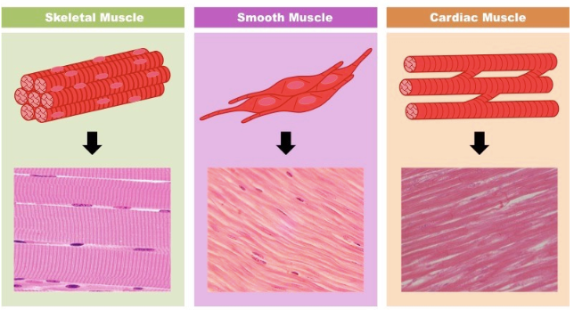
• Smooth Muscles * Blood vessels and hollow organs * Involuntary control * Not striated * Single nucleus • Cardiac Muscles * Heart muscle * Involuntary control * Striated * Single nucleus • Skeletal Muscle * Movement * Voluntary control * Striated and tendons * Multinucleated
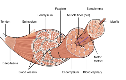
1.2 Muscular system Structure of a skeletal muscle
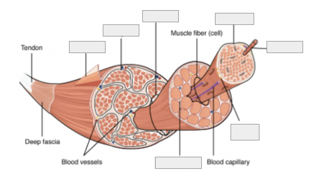
• Epimysium - layer that covers entire muscle • Perimysium - surrounds bundles of muscle fibres • Muscle fiber (fascicles) - strand of muscle • Endomysium - surrounds individual fibres • Sarcomere - repeating unit in myofibril, where contraction happens • motor nueron • Myofibril - sub strand of fibre • Actin - Thin filament • Myosin - Thick filament
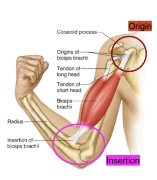
1,2 Muscular system How to describe muscle locations
Two descriptors of the specific location of a muscle are based on where it attaches to bone, where it originates and where it inserts (ends) * Origin - the muscle tendon attachment point that is stationary with contraction of the muscle. Usually proximal * Insertion - the muscle tendon attachment point that moves with contraction of the muscle. Usually distal
4.1 Neuromuscular Function The motor unit
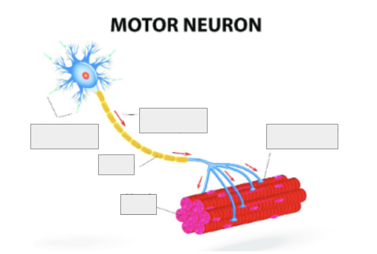
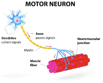
A motor unit is made up of a motor neuron and all of the skeletal muscle fibers innervated by the neuron’s axon terminals To include: Dendrite, cell body, nucleus, axon, motor end plate, synapse, muscle
4.1 Neuromuscular Function How the signal from the nerve get to the muscle
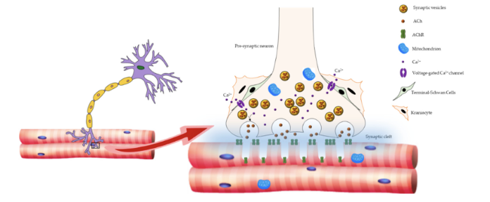
• neurotransmitters The role of neurotransmitters in stimulating skeletal muscle contraction: • Neurotransmitters are chemical messengers • They are responsible for transferring the electrical signal from the neuron, across the synapse as a chemical messenger, to the muscle cell • Two specific NT are used in stimulating a muscle contraction: Acetylcholine: –>Produced in neuron and triggers a receptor on the muscle Cholinesterase: –> Removes acetylcholine from the muscle receptor –> Breaks acetylcholine down into acetyl and choline
4.1 Neuromuscular Function The 7 steps of a neurotransmitter stimulating a skeletal muscle contraction:
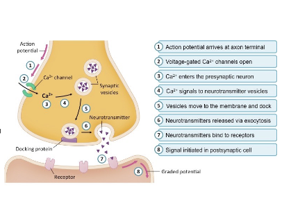
• Choline and acetyl combine to create acetylcholine in the axon terminal (end of neuron) • Acetylcholine is stored in vesicles • A nerve impulse reaches the end of the neuron causing acetylcholine to be released via exocytosis into the synaptic cleft • Acetylcholine travels across the synaptic cleft • Acetylcholine binds to a receptor on the muscle fiber and triggers depolarization of the muscle cell • Depolarization releases calcium ions (Ca2+) which is used to trigger a muscle contraction • Cholinesterase removes acetylcholine from the receptor to stop the signal for a contraction • Choline is recycled by the neuron to be used again (step 1)
4.1 Neuromuscular Function Sliding filament theory (what, structures involved, steps (8), end result)
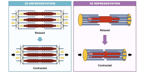
• during muscle contraction ACTIN slides over MYOSIN Structures involved: Adenosine triphosphate (ATP) is a high energy molecule that functions as an immediate power source for cells When ATP is hydrolysed (to form ADP + Pi), the energy stored in the terminal phosphate bond is released for use by the cell steps: 1. Action potential in a motor neuron triggers the release of Ca2+ ions from the sarcoplasmic reticulum 2. Calcium ions bind to troponin (on actin) and cause tropomyosin to move, exposing binding sites for the myosin heads 3. ATP binds to myosin head 4. ATP hydrolysis (ATP → ADP + P) causes the myosin heads to change orientation 5. Myosin heads binds to the actin filament creating a cross-bridge 6. ADP and P release resulting in ENERGY released causing the myosin head to move the actin filaments towards the center of the sarcomere (power stroke) 7. ATP is able to re-attach to myosin head breaking the cross bridge 8. Cycle is able to restart end result: The sliding of actin along myosin therefore shortens the sarcomere, causing muscle contraction –> this decreases the length of the h Zone and I band of the sarcomere * A band remains the same length * Actin and myosin do not change in length. They simply overlap each other more causing shortening
4.1 Neuromuscular Funtion Types of muscle fibres
• Type I - Slow twitch • Type IIa - Fast twitch oxidative • Type IIb - fast twitch glycolytic
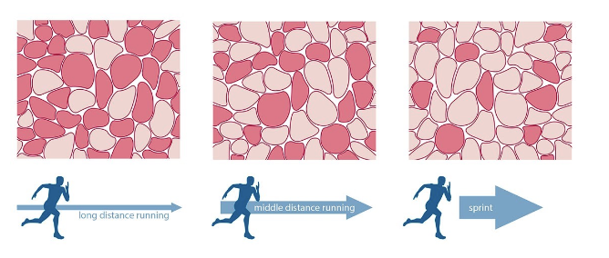
structure (6) and function (4) of slow twitch muscle fibre
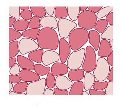
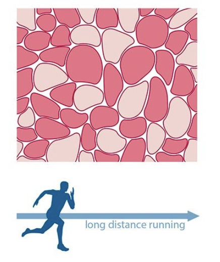
Type I - Slow twitch Structure: * have a large amount of mitochondria and many blood vessels * Typically red in color due to the dense supply of capillaries * Small sized motor unit * Fuel is Triglycerides (fatty acids) - has storages * High myoglobin - part of muscle that use oxygen * Smaller amounts of sarcoplasmic reticulum Function: * Endurance focused * Slow nerve contraction speed –> Slow to contract –> Fatigue resistant –> Small force generated * Slow twitch fibers use oxygen for aerobic respiration * Prevalent in endurance athletes, such as marathon runners
structure (7) and function (7) of type IIa muscle fibre
Type IIa - Slow twitch oxidative Structure: * Respire anaerobically * High mitochondria and have medium blood vessels density * Typically lighter in color (white) * Large size of motor unit * High in Glycogen and Creatine as fuel source and stores * High myoglobin - part of muscle that use oxygen * Larger amount of sarcoplasmic reticulum Function: * Strength focused * Faster nerve contraction speed * Quick to contract * Intermediate Fatigue - produce lactic acid * Larger force generated * Do not use oxygen, anaerobic respiration * Prevalent in strength athletes, middle distance runners, cycling
s structure (7) and function (7) of type IIb muscle fibre
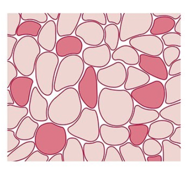
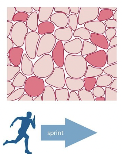
Structure: * Respire anaerobically * Low mitochondria and have low blood vessels density * Typically lighter in color (white) * Largest size of motor unit * High in Glycogen and Creatine as fuel source and stores * Low myoglobin - part of muscle that use oxygen * Larger amount of sarcoplasmic reticulum Function: * Explosive power focused * Fastest nerve contraction speed * Quickest to contract * High Fatigue - produce lactic acid * Largest force generated * Do not use oxygen, anaerobic respiration * Prevalent in explosive athletes; sprinters (100m)/jumpers/ weight lifters
4.2 Joint and movement type Types of movement of synovial joints Forward and backwards movement
• Flexion - decreasing the angle at a joint (bicep curl) • Extension - increasing the angle at a joint (straightening your leg at knee)
difference between type IIa and type IIb
Type IIa - Fast twitch oxidative Structure: * Respire anaerobically * High mitochondria and have medium blood vessels density * Typically lighter in color (white) * Large size of motor unit * High in Glycogen and Creatine as fuel source and stores * High myoglobin - part of muscle that use oxygen * Larger amount of sarcoplasmic reticulum Function: * Strength focused * Faster nerve contraction speed * Quick to contract * Intermediate Fatigue - produce lactic acid * Larger force generated * Do not use oxygen, anaerobic respiration * Prevalent in strength athletes, middle distance runners, cycling Type IIb - fast twitch glycolytic Structure: * Respire anaerobically * Low mitochondria and have low blood vessels density * Typically lighter in color (white) * Largest size of motor unit * High in Glycogen and Creatine as fuel source and stores * Low myoglobin - part of muscle that use oxygen * Larger amount of sarcoplasmic reticulum Function: * Explosive power focused * Fastest nerve contraction speed * Quickest to contract * High Fatigue - produce lactic acid * Largest force generated * Do not use oxygen, anaerobic respiration * Prevalent in explosive athletes; sprinters (100m)/jumpers/ weight lifters
4.2 Joint and movement type Side to side type of movements of synovial joints
• abduction - moving away from the midline of your body (lateral raise of your arm) • adduction - moving towards the midline of your body (bringing your legs together while standing)
4.2 Joint and movement type Up and down types of movement of synovial joints
• elevation - raising a body part verticaly ex. shrugging your shoulders • depression - lowering a body part vertically ex. opening your jaw
4.2 Joint and movement type other ways to describe movement of synovial joints (8)
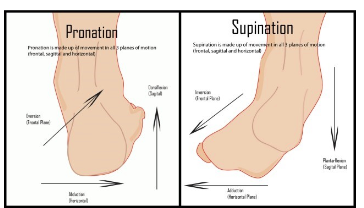
• Rotation (Internal/External) - spinning around a fixed axis (looking right or left) • Circumduction - moving a limb in a circle (arm circles) • Pronation - palms down OR 3 axis movement of ankle • Supination - palms up 3 axis movement of ankle • Dorsiflexion - raising your toes • Plantar flexion - pointing your toes • Eversion - outward turn of ankle • Inversion - inward turn of ankle