ChemPath: Clinical Chemistry CPC
No description yet. This curated deck is ready for daily study.
Card preview (24)
Describe the effect of hypokalaemia on the myocardium.
Increases the myocardial irritability.
Describe the cardiac consequences of plasma potassium being too high or too low.
• Too low - ventricular fibrillation • Too high - asystole (ultimate stable rhythm)
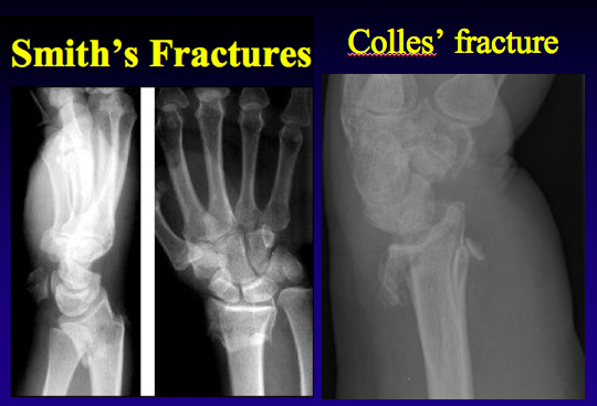
What is the difference between a Colles’ fracture and a Smith’s fracture?
• Colles’ - fracture caused by falling on an outstretched hand. The radial head will be displaced backwards (away from the palm). • Smith’s - fracture caused by falling on a flexed wrist. The radial head will be displaced forwards (towards the palm)
What is a Pott’s fracture?
Ankle fracture involving the tibia and fibula
What would you expect to see on the urine dipstick of someone with subacute bacterial endocarditis?
Microscopic haematuria
What is the physiological role of PTHrP?
• Our genome encodes a gene for PTHrP • This is important in foetal life because it allows us to steal calcium from our mother to help form our skeleton NOTE: PTHrP is also produced by the lactacting breast NOTE: PTHrP stimulates cancer cells to invade bone
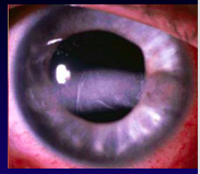
Name and describe an eye sign of hypercalcaemia.
• Band keratopathy - calcium deposition across the front of the eye • It is a feature of chronic hypercalcaemia (i.e. it will not be caused by hypercalcaemia of malignancy)
What is a key difference between calcium stones and urate stones?
Calcium stones are radio-opaque Urate stones are radiolucent
List some complications of hypercalcaemia.
• Renal stones • Pancreatitis • Peptic ulcer disease • Skeletal changes (osteitis fibrosa cystica)
List some risk factors for hypercalcaemia.
Family history Dehydration Hyperparathyroidism
Which bacterium has a predilection to infect urinary tract stones?
Proteus mirabilis
What are the main investigations used for urinary tract stones?
• CT-KUB • Stone analysis • Urine and serum biochemistry
What are some management options for urinary tract stones?
• Lithotripsy • Cystoscopy • Lithotomy
How can urinary tract stones be prevented?
• Drink more water • Treat hypercalciuria (thiazides) • Treat hypercalcaemia NOTE: loop diuretics increase urine calcium
At what point would you use emergency management of hypercalcaemia?
When serum calcium > 3 mmol/L or very unwell (e.g. dehydrated, confused, drowsy, seizures)
Outline the emergency management of hypercalcaemia.
• IV access • Insert catheter • 3-6 L 0.9% saline over 24 hours • The first litre should be given quickly (over 1 hour) to correct dehydration • Elderly patients should also be given furosemide (to prevent pulmonary oedema)
Which other drug may be used under desperate circumstances when managing hypercalcaemia?
• Pamidronate (IV) • Good at treating bone pain but takes at least 1 week to start working and gets incorporated into bone for a very long time
In which group of patients would you used dextrose rather than saline?
Liver failure - they have a tendency to retain salt
Outline the treatment of non-emergency hypercalcaemia.
• Keep well hydrated • Avoid thiazides (they reduce hypercalciuria but they increase plasma calcium concentration) • Surgery
What is minimally invasive parathyroidectomy?
• A technetium sesta MIBI scan shows a hyperactive parathyroid • An USS is also performed and if the results of the sesta MIBI and USS are concordant, the whole neck does not need to be opened • If they are not concordant, the surgeon will need to view all four glands and take out the largest one
What feature may you see on an X-ray of the hands in a patient with primary hyperparathyroidism?
Cystic changes in the radial aspect
What is a characteristic histological feature of long-standing undiagnosed hyperparathyroidism?
Brown tumours - they are multinucleated giant cells in the bone. The giant cells are activated osteoclasts.
What is the mainstay of treatment of sarcoidosis?
Steroids
What is the histological hallmark of sarcoidosis?
Non-caseating granulomas