Unit 3: Exercise Physiology - 2.1 Ventilation
No description yet. This curated deck is ready for daily study.
Card preview (16)
Principal structures of the ventilatory system (9)
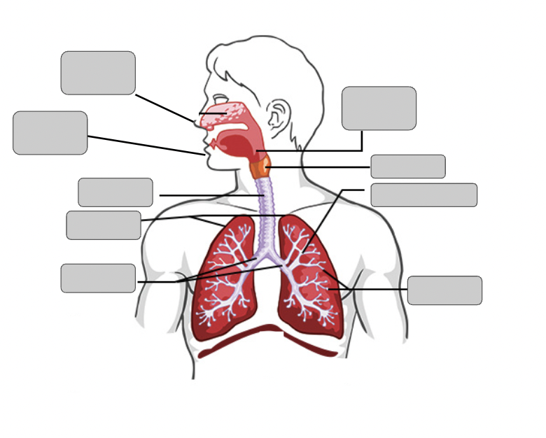
• nose • mouth • pharynx • larynx • trachea • bronchi • bronchioles • lungs • alveoli
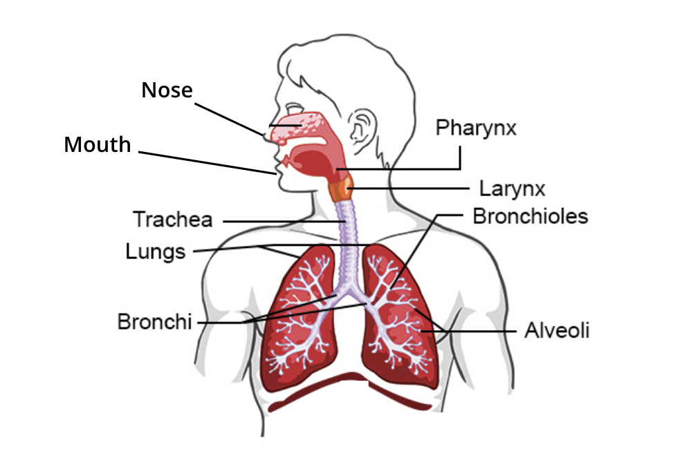
Advantages of the structures for ventilation (5)
• All about making it easier for air to get in but hard for other particles to get in • Nose: Humidifies the air entering and hair filters particles • Pharynx: low resistance path for airflow into the larynx • Larynx: In addition to its function as the ‘voice box’ it also protects the trachea from invasion of foods and fluids. • Trachea: Mucus coating that traps pathogens and dirt. Cilia hairs move phlegm out of trachea
Process of ventilation (6)
Process of ventilation: 1. Air enters the lungs because muscles increase the volume of the thoracic cavity. Air enters to fill this space, expanding your lungs. 2. Air exits your lungs because muscles decrease the volume of the thoracic cavity. Air is pushed out of your lungs. • Gasses will move from a region of high pressure to a region of lower pressure When the volume of the thoracic cavity increases, pressure in the thorax decreases –> When the pressure in the chest is less than the atmospheric pressure, air will move into the lungs (inspiration) When the volume of the thoracic cavity decreases, pressure in the thorax increases –> When the pressure in the chest is greater than the atmospheric pressure, air will move out of the lungs (expiration)
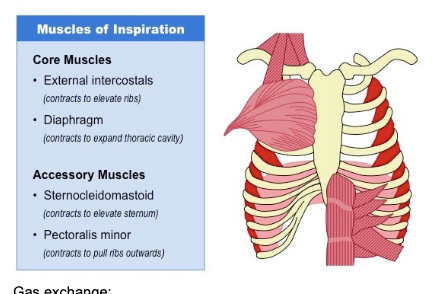
Inhalation steps
Increasing the volume of thoracic cavity • Diaphragm muscles contract, causing the diaphragm to flatten and increase the volume of the thoracic cavity • External intercostals contract, pulling ribs upwards and outwards (expanding chest) • Additional muscle groups may help pull the ribs up and out (e.g. sternocleidomastoid and pectoralis minor) muscles involved: 1. core musckes: * external intercostals (contract to elevate ribs) * diaphragm (contracts to expand thoracic cavity) • Accesory muscles: * sternocleidomastoid (Contracts to elevate sternum) * pectoralis minor (contracts to pull ribs outwards)
Exhalation steps
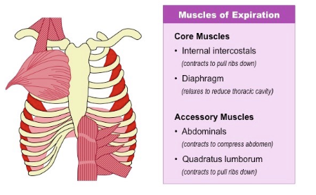
Decreasing the volume of thoracic cavity • Diaphragm muscles relax, causing the diaphragm to curve upwards and reduce the volume of the thoracic cavity • Internal intercostal muscles contract, pulling ribs inwards and downwards (reducing breadth of chest) • Additional muscle groups may help pull the ribs downwards (e.g. quadratus lumborum) muscles involved: 1. core muscles: * internal intercostal (contracts to pull ribs down) * diaphragm (relaxes to reduce thoracic cavity) • accesory muscles: * abdominals (contracts to compress abdomen) * qudratus lumborum (contract to pull ribs down)
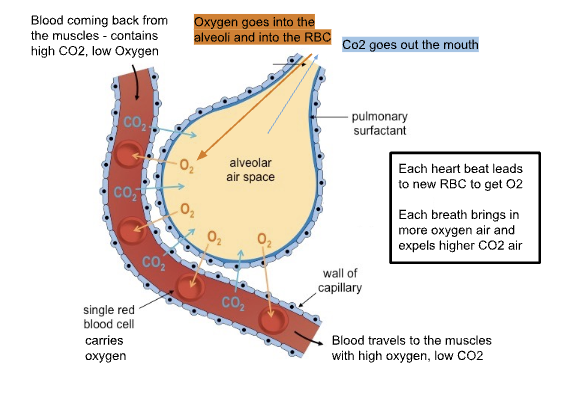
Gas exhange process:
The final step, all about getting oxygen to red blood cells from the alveoli, and carbon dioxide into the alveoli from the RBC. • happens at the alveoli
Role of hemoglobin in oxygen transportation (5)
• Hemoglobin is a protein found INSIDE red blood cells. • Hemoglobin is the molecule that carries oxygen. • Red blood cells are able to carry oxygen because they have hemoglobin. –> You have ~250 million hemoglobin in each RBC • Hemoglobin contains a central Iron ion and can hold 8 oxygen atoms molecules. • When oxygen is attached to hemoglobin it is called oxyhemoglobin
VO2 definition
volume of oxygen absorbed by the body per minute
VO2 max meaning
the maximum amount of oxygen your body could possible absorb per minute (trainable)
Frequency definition
breaths per minute
spirometry
measures the amount and rate of air a person breathes in order to diagnose illness or determine progress in treatment
Response to exercise (what -2, why-4)
• Increase ventilation rate (a greater frequency of breaths allows for a more continuous exchange of gasses) • Increase tidal volume (increasing the volume of air taken in and out per breath allows for more air in the lungs to be exchanged because: * Ventilation in humans changes in response to levels of physical activity, as the body’s energy demands are increased * ATP production (via cellular respiration) produces carbon dioxide as a waste product (and may consume oxygen aerobically) * Changes in blood CO2 levels are detected by chemosensors in the walls of the arteries which send signals to the brainstem * As exercise intensity increases, so does the demand for gas exchange, leading to an increase in levels of ventilation
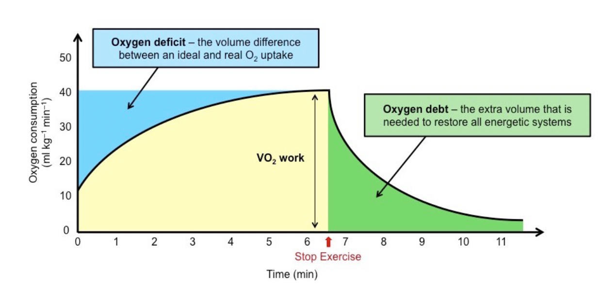
Oxygen debt Why do we need to continue to breathe heavily once we stop exercising? (4)
• If our body’s demand for oxygen is being met by our VO2 then everything is okay • If the body needs more oxygen than what we are able to take in, we go into oxygen debt. (aerobic vs anaerobic) • We need to pay this debt after exercise stops to reset our body • Excess Post-exercise Oxygen Consumption (EPOC)
How does our body nervously control breathing (7)
• The brain stem has sections called the medulla oblongata and pons that are in charge of respiration • They have nerves that go to the inspiratory muscles and expiratory muscles to control breathing. During exercise they send signals to ventilation muscles to increase frequency and volume of ventilation. –> Increase Frequency is caused by nerves triggering more frequent expiration through the expiratory muscles. Changing from passive expiration to active control. –> Increased volume is caused by nerves triggering inspiratory muscles to contract more forcefully. Triggers more accessory muscles to help. • Stretch receptors in the lungs stop us from being able to over-inflate our lungs. Hering-Breuer Reflex –> Stretch Receptors in the lungs are triggered when they are stretched too much because of inflation of the lung —> They send a signal to the brain’s respiratory center to shorten the length of inhalation • Proprioceptors relay information to the respiratory center regarding action of muscles / joint receptors • Chemoreceptors relay information to the respiratory center regarding lower pH or O2 / higher CO2 levels
How does our body chemically control breathing: (5)
• Ventilation is chemically regulated by blood acidity levels/ pH • Blood acidity levels increase/drops due to an increase/decrease in carbon dioxide levels. CO2 levels trigger breathing not O2 • Blood acidity levels are detected by chemoreceptors in the brain and arteries. • Medulla oblongata (respiratory control center) receives information from receptors • Increased blood acidity increases the rate of ventilation
ventilation and exercise the bodies response to exercise
• Increase ventilation rate (a greater frequency of breaths allows for a more continuous exchange of gasses) • Increase tidal volume (increasing the volume of air taken in and out per breath allows for more air in the lungs to be exchanged