2 - Cells
No description yet. This curated deck is ready for daily study.
Card preview (24)
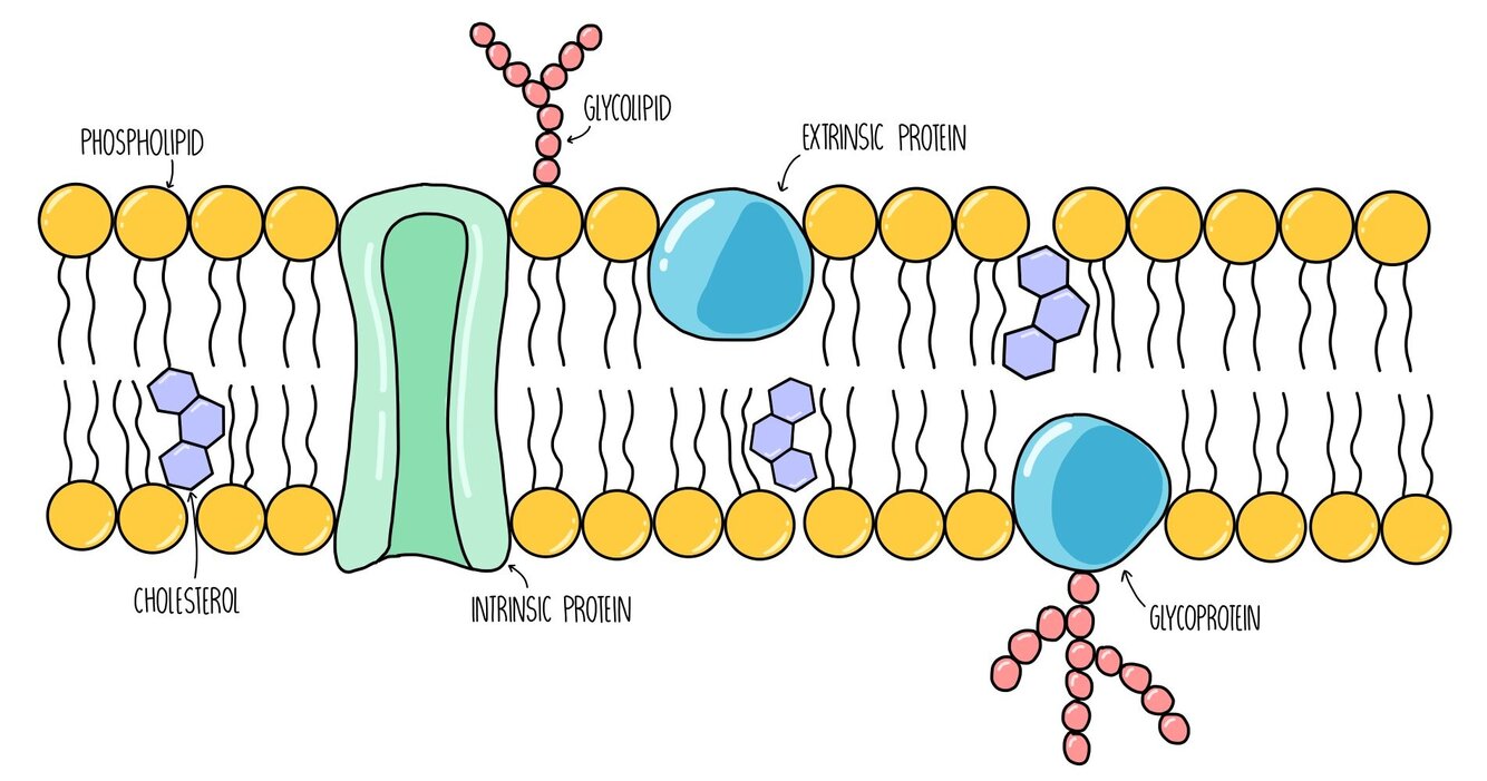
Describe the function of the cell surface membrane:
allows for exchange of substances
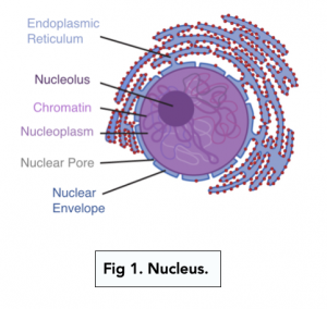
Describe the structure of the nucleus:
-nuclear envelope (double membrane) with many pores -nucleoplasm -chromatin (diffused, uncondensed material that makes up chromosomes) -nucleolus (may be more than one)
Give some functions of the nucleus:
-controlling the production of m/tRNA for protein synthesis -holding genetic material in the form of linear DNA -to manufacture ribosomal RNA and ribosomes in the nucleolus
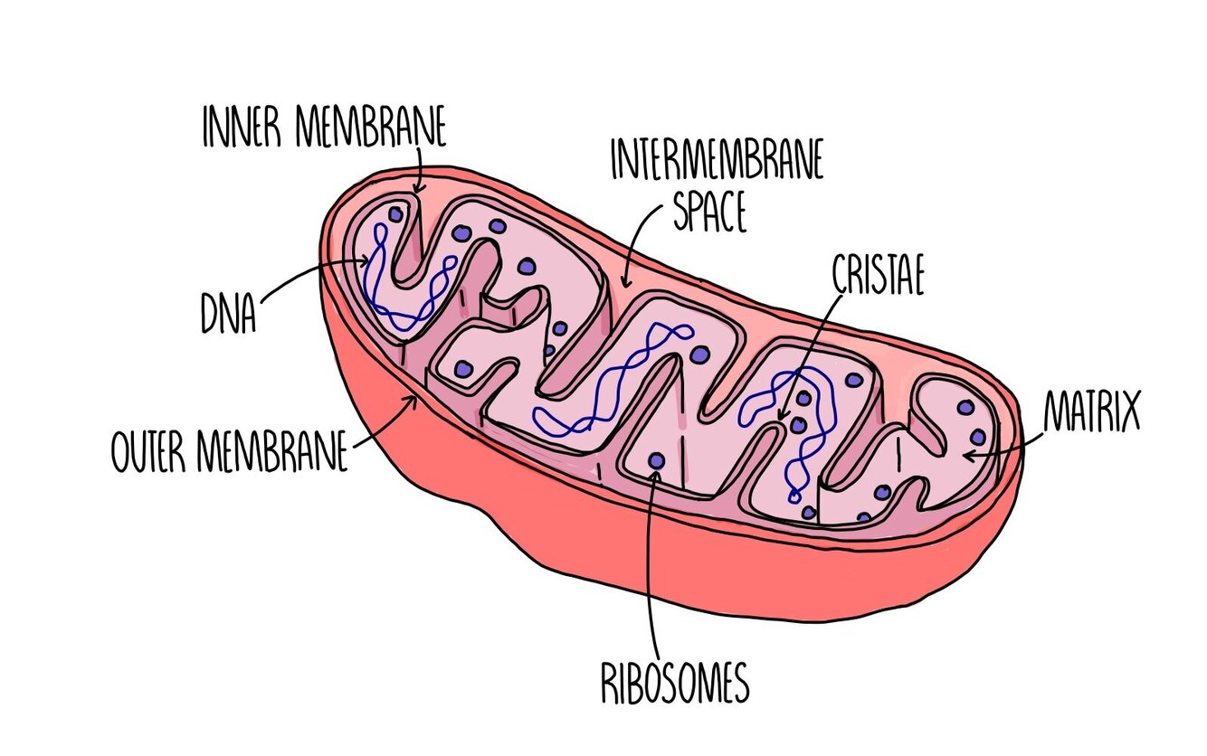
Name the features of a mitochondrion:
-double membrane (inner membrane has folds known as cristae) -viscous fluid inside is the matrix, and has the enzymes needed for respiration -ribosomes Can also have mitochondrial DNA
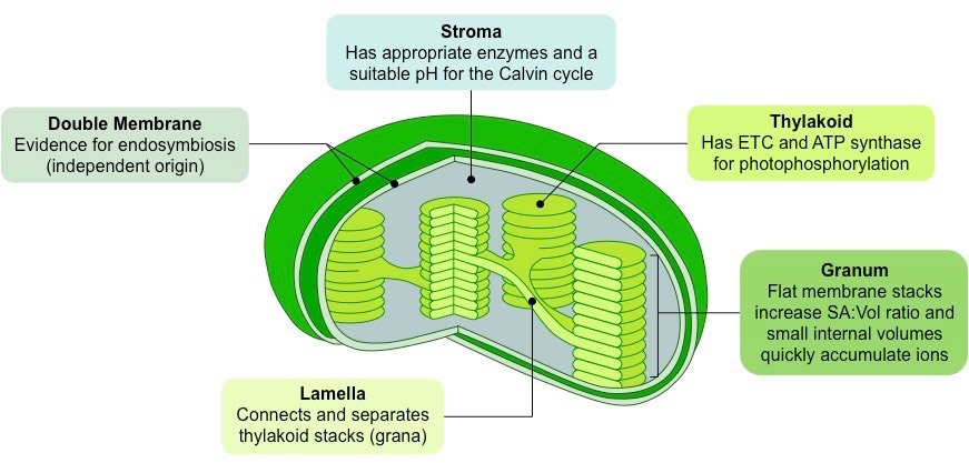
What are some features of chloroplasts?
-chloroplast envelope (double membrane) -grana (stacks of thylakoid discs containing chlorophyll) -stroma (fluid) -starch grains -ribosomes Light-dependent reactions occur in thylakoids, independent in stroma
Give a feature of chloroplasts that allow proteins to be synthesised inside of them, and explain how it is different to the rest of the cell:
-they have DNA in them (similar to prokaryotic DNA, not associated with proteins and circular) -they have 70S ribosomes, which are smaller than the 80S cytoplasmic ribosomes
Describe the structure of the Golgi apparatus:
-flattened sacs of single membranes called cisternae (similar to SER) -Golgi vesicles forming by pinching off parts of the cisternae cis face is closest to ER, trans is furthest (medial is inbetween)
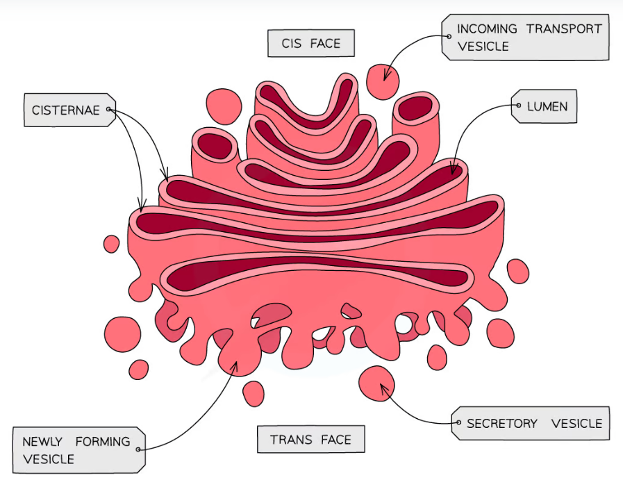
Explain how molecules get used by the Golgi apparatus:
-incoming proteins/lipids are in vesicles from the ER -modified and processed (eg adding carb. to make glycoprotein, glycosylation) -molecules then leave in Golgi vesicles to be secreted Molecules secreted could be enzymes like in the pancreas, or carbohydrates like in plants for cell walls
What is a lysosome?
a specialist form of a vesicle that contains hydrolytic enzymes to break down biological molecules They are made when the vesicles from the GA contain enzymes
Give some of the main functions of lysosomes:
-to hydrolyse material ingested by phagocytic cells -to secrete enzymes to the outside (exocytosis) -digesting worn-out organelles to recycle their materials -breaking down cells after death (autolysis)
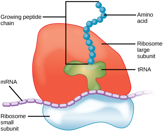
Describe the structure of a ribosome:
-composed of a large sub-unit and a small sub-unit -both contain rRNA and proteins
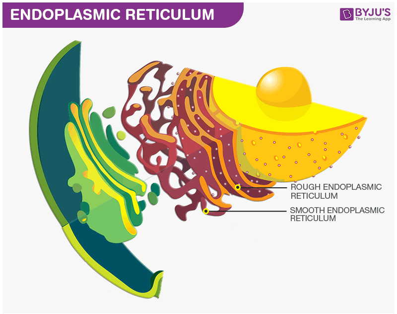
What is the endoplasmic reticulum?
-a system of flat sheet-like membranes (cisternae) RER -membranes are continuous with nuclear envelope and is covered in ribosomes SER -no ribosomes, and is involved in the synthesis/storage/processing of lipids/carbs/steroids
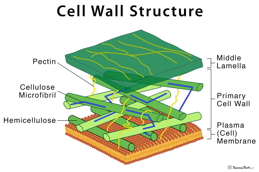
What is the structure of the cell wall?
-middle lamella that marks as a boundary between cell walls, cementing them together -many polysaccharides including microfibrils of cellulose
How do cell walls differ between different types of organisms?
-in plants, it is made of cellulose -in algae, it is made of cellulose or glycoproteins (or both) -in fungi, it is made of chitin -in bacteria, it is made of murein (aka peptidoglycan)
Describe the structure of a vacuole and how it contributes to its function:
-single membrane enclosing the cell sap is known as the tonoplast -makes cells turgid for support by drawing water into the cell -can act as a temporary food source (contains sugars + amino acids)
Why are arteries and veins considered organs, but not capillaries?
-arteries/veins contain multiple types of tissue -capillaries only have epithelial tissue
How is the DNA different in prokaryotic cells?
prokaryotes have a single circular DNA molecule that is free to move about the cytoplasm (not inside a nucleus), and is not associated with proteins The proteins specified are histones - a type of protein that allows for chromatin to be condensed and packed into chromosomes
Explain why a virus might only be able to infect one type of cell:
-the virus surface has attachment proteins -its shape is complementary to the receptor proteins found on the cells -only those types of cells will have the complementary receptor protein
What is the formula for magnification?
image size/actual size
Why is the solution buffered for storage before cell fractionation?
to stop large fluctuations in pH, which prevents the structure of organelles or enzymes from being altered
How can you obtain a cell-free solution after homogenisation and filtration?
-centrifuge the sample at a low speed -denser cells and cellular debris form a pellet at the bottom of the tube -decant the supernatant into a separate tube
Explain the principles behind how a TEM works:
-electrons pass through thin specimen -denser parts absorb more electrons and will appear darker -very short wavelength so higher resolution achieved
Give some cons of using electron microscopes:
-specimen must be dead (either due to heavy metal stains, or because it is a very thin slice for TEMs, or because it is in a vacuum) -can’t have true colour images -lengthy sample preparation
How are specimens measured under microscopes?
-eyepiece graticule is calibrated using a slide with a stage micrometer on it -the length of each EPG unit is calculated using a ratio with the stage micrometer, so then specimens can be measured accurately