Theme 4: Cardiovascular Biology During Stress and Disease
No description yet. This curated deck is ready for daily study.
Card preview (24)
What are the main categories of birth defects?
Structural: • Abnormalities in the development of body parts • Not amenable to treatment, but may be correctable by surgery Functional: • Abnormalities in the systems that run the body • Vary in severity from inconsequential (hammer toe) to life-threatening (heart defects) • Includes metabolic disorders (phenylketonuria) and degenerative diseases (muscular dystrophy) • May be treatable in some cases
Give some statistics about congenital heart defects.
• Heart defects are the most common class of structural birth defects • CHD affects ~1% of all live births, on average 13 per day in the UK • Despite modern surgery, still the biggest cause of infant mortality and morbidity • May affect up to 10% of still births and spontaneous terminations
How are congenital heart defects detected?
• Phenotyping is a full investigation of the nature and severity of defects • This is done via 2D and 3D ultrasound from 20 weeks of pregnancy • After birth, phenotyping may continue using Magnetic Resonance Imaging (MRI) or X-Ray/CT scan
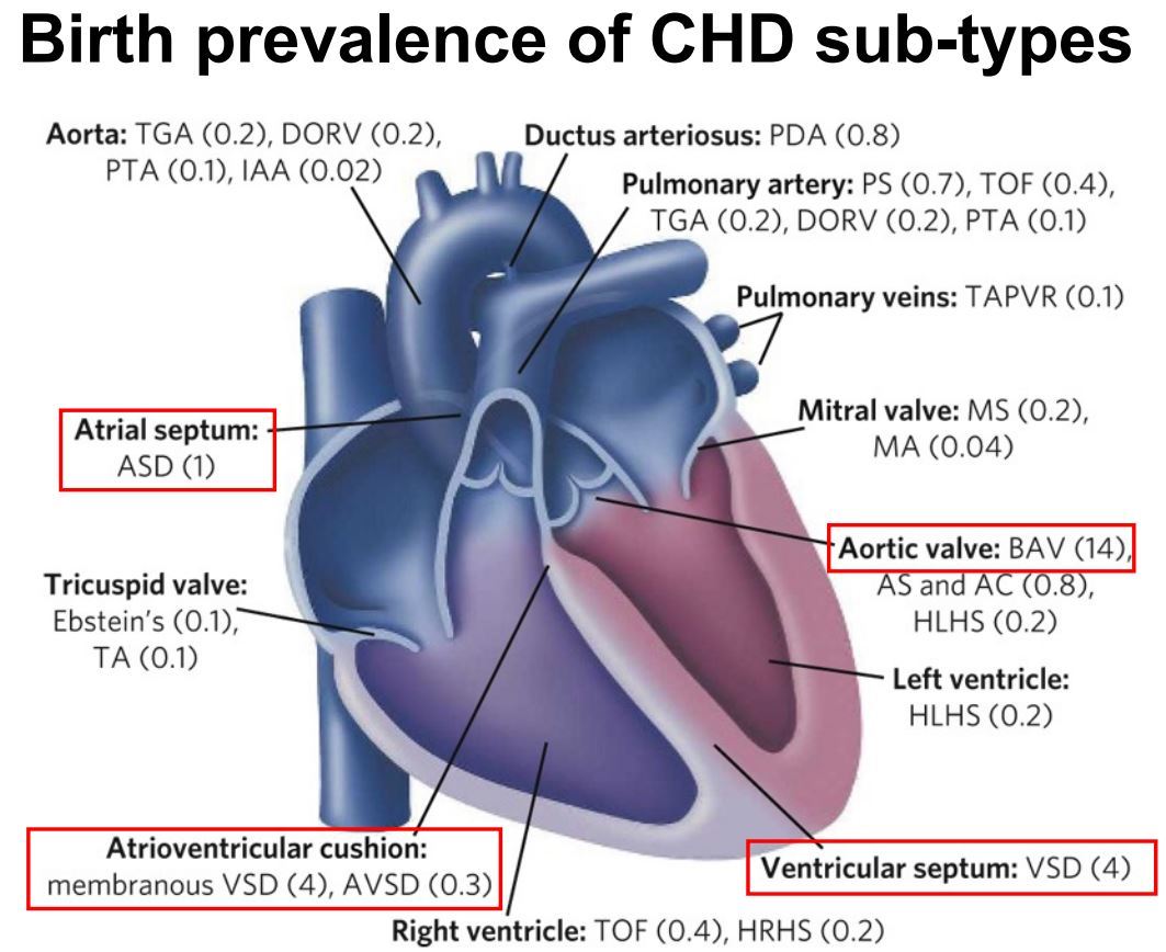
What are the most common types of congenital heart defect?
The most common is a bicuspid aortic valve, which is not usually a problem until later life when there is a higher risk of calcification.
What percentage of congenital heart defects have a simple genetic cause?
About 30%
What are the main types of inherited congenital heart defect?
• Chromosomal abnormalities • Copy number variation (CNV) • Small nucleotide polymorphisms (SNP)
Describe what chromosomal abnormalities are and how they can be diagnosed.
• These can involve duplication or absence of an entire chromosome • Alternatively they can be insertion, deletion, duplication, translocation or inversion of a large portion of a chromosome (many Mb) • The best known example is Down syndrome (trisomy 21) where 40% of patients have CHD • They can be diagnosed by isolating cells from the embryo or placenta and looking at the karyotype, fluorescence in situ hybridization (FISH) or PCR -> The latest techniques enable embryonic cells to be isolated from a sample of the mother’s blood from 9-12 weeks
Describe what copy number variations (CNVs) are and how they can be diagnosed.
• These are duplications or deletions of small regions of chromosome, typically 1-18 kb • These can be detected by fluorescence in situ hybridization (FISH), array comparative genomic hybridization (aCGH) or next generation sequencing • Typically cause deletion or duplication of one entire gene
How does searching for SNPs that cause congenital heart defects work?
• The human genome has ~3 billion base pairs • The reference genome is derived from the DNA of 13 anonymous donors (USA) • Any particular individual has ~3 million SNPs that vary from the reference genome • Exome sequencing is often used: • Exome sequencing requires exon isolation (hybridisation) followed by next generation sequencing • This is rapid but only covers <90% of the exome, and misses non-coding variation • An exome dataset has ~60k variants, with ~100 “private” or unique to that individual/family • It then remains a task to identify which of these variants is causative of the disease (see table)
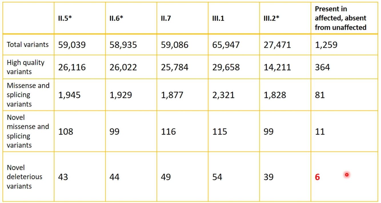
How do we distinguish between natural variation (benign) and disease-causing variation (pathogenic)?
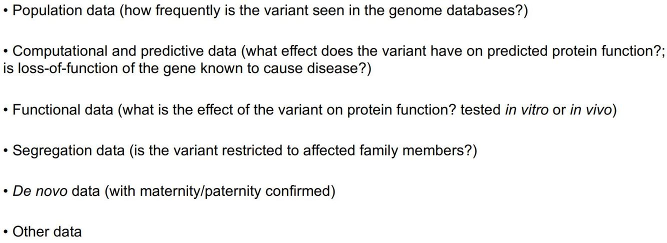
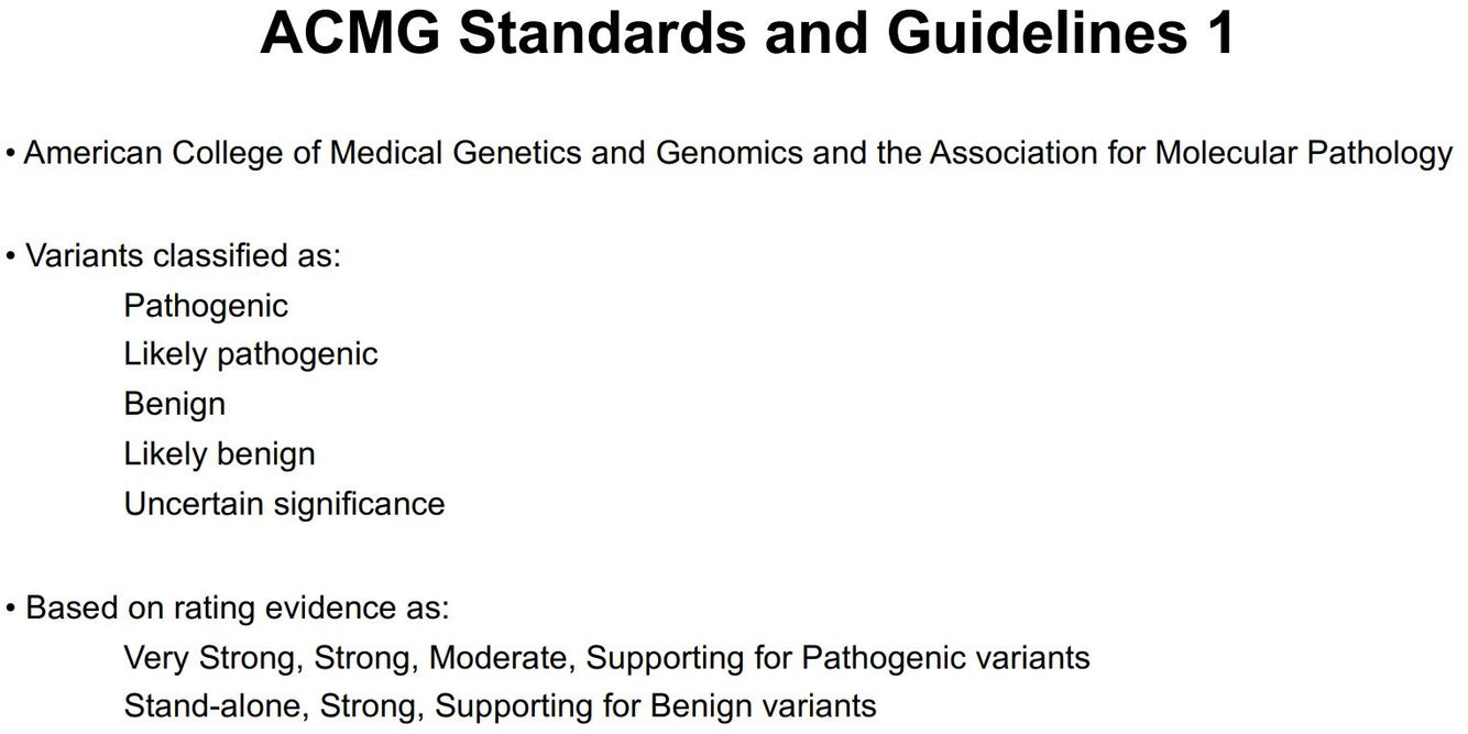
What standards are used for classifying how pathogenic a variant is pathogenic?
What are the main limitations of exome sequencing and what is the alternative?
• It doesn’t cover ~10% of exons • It misses variation in gene regulatory regions that might be causing defects Whole genome sequencing is the alternative. It is now cheap enough to use clinically (around £1000 per sample), but it generates huge amounts of data, which become difficult to store.
What are some environmental risk factors for congenital heart defects?
• Infectious diseases (e.g. Rubella and Zika viruses) • Environmental teratogens (e.g. hypoxia, hyperthermia) • Maternal nutritional deficiencies (e.g. Vitamin B3, iron) • Maternal non-communicable diseases (e.g. pre-gestational diabetes) • Maternal genetic conditions (e.g. phenylketonuria) • Teratogenic therapeutic drugs (e.g. Thalidomide, Roaccutane)
What animal models can be used for studying congenital heart defects?
• Mouse embryos • However, they do not always respond in the same was as humans (e.g. they do not respond to thalidomide)
What type of epidemiological study is usually used to study congenital heart defects?
Retrospective observational case-control studies
What are the limits of epidemiological study of congenital heart defects?
• All the key events of human embryonic heart development occur before week 8 of gestation. Therefore, the mother may not accurately remember her exposure to risk factors when the birth defect is diagnosed. • Likewise, clinical workup of maternal physiological parameters when the birth defect is first detected will not necessarily reflect her conditions during weeks 1-8. • Comprehensive maternal histories often not recorded • Can take decades to satisfactorily test if the association is real (e.g. folic acid) • Can take years before risk factor and/or corrective therapy becomes widely known (Valproate for epilepsy)
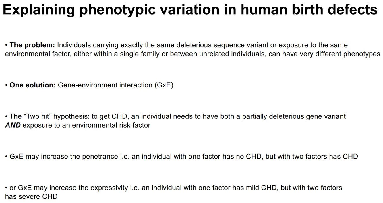
Explain the importance of gene-environment interaction (GxE) in congenital heart defects.
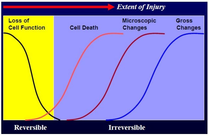
What is the difference between myocardial ischaemia and infarction?
Infarction is when the cells die.
Define ischaemia.
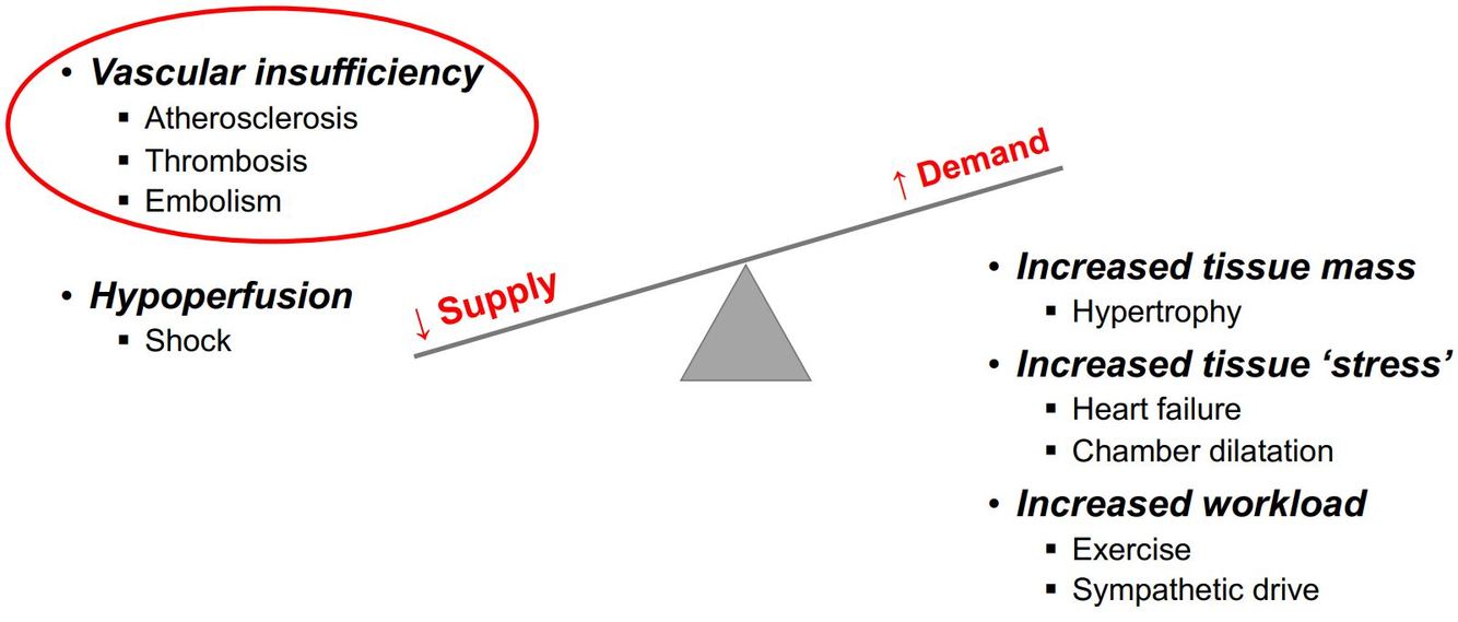
• Ischaemia occurs when blood supply to a tissue is inadequate to meet the tissue’s demand. • There are 3 main components: • Hypoxia • Insufficiency of metabolic substrates • Accumulation of metabolic waste
Name some ways in which supply and demand of the heart may become unbalanced, leading to ischaemia.
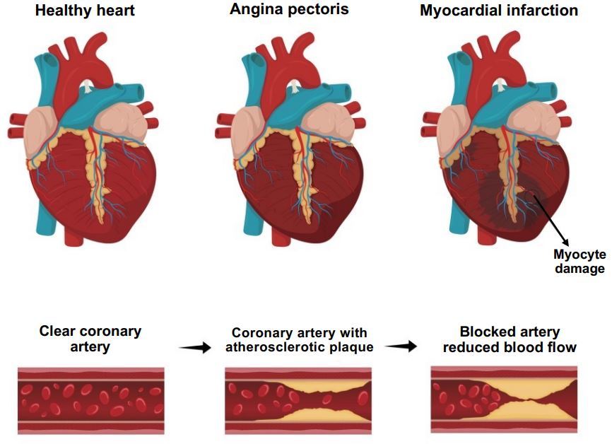
Compare angina and myocardial infarction.
• Angina (stable, unstable) • Chest pain caused by myocardial ischaemia • Usually due to atherosclerosis • Myocardial infarction • Complete occlusion of coronary flow • Plaque rupture with occlusive thrombus
How is a myocardial infarction diagnosed?
• The gold-standard marker of myocardial injury is blood test (ELISA) for cardiac troponin (cTn) I or T. • This is because troponin is usually found inside cardiac myocytes.
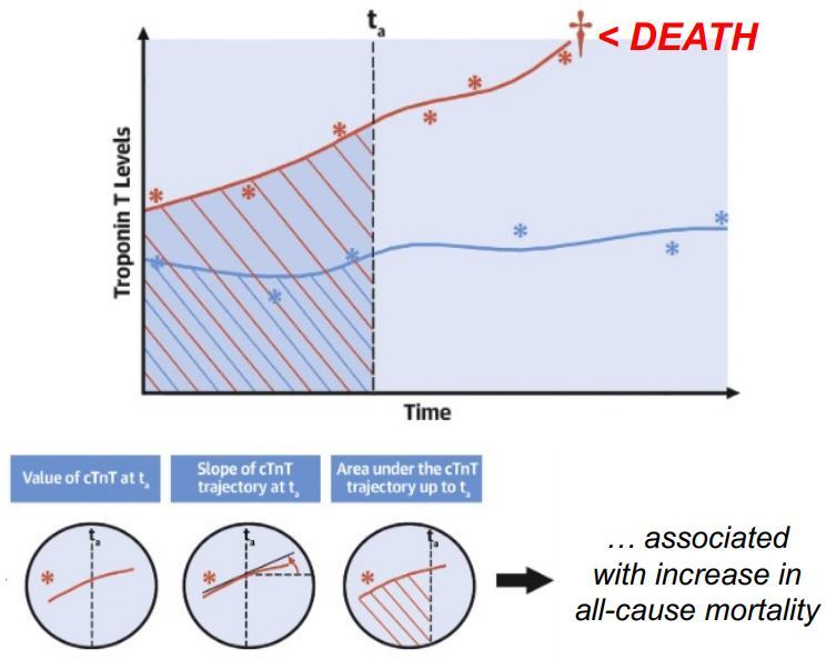
What is an increased blood cTnT (cardiac troponin T) associated with? Give experimental evidence.
• It is not only an indicator of myocardial infarction, but it also appears to be associated with all-cause mortality • (Chesnaye, 2022): • It has previous been shown that cardiac troponin T (cTnT) is associated with mortality in chronic kidney disease (CKD). • Studied 176 patients with stage 4-5 CKD aged ≥65 years and not on dialysis • Took cTnT measurements over a median follow-up of 2.4 years • Found that 3 factors were associated with all-cause mortality@ • Longitudinally measured cTnT • Slope of the cTnT trajectory • Area under the cTnT trajectory
How does extreme exercise influence cardiac troponin T (cTnT)?
• Extreme exercise has been shown to lead to increases in troponin T • 2/3rds of individuals after a marathon gave a positive blood troponin test