2. Mediastinum
No description yet. This curated deck is ready for daily study.
Card preview (10)
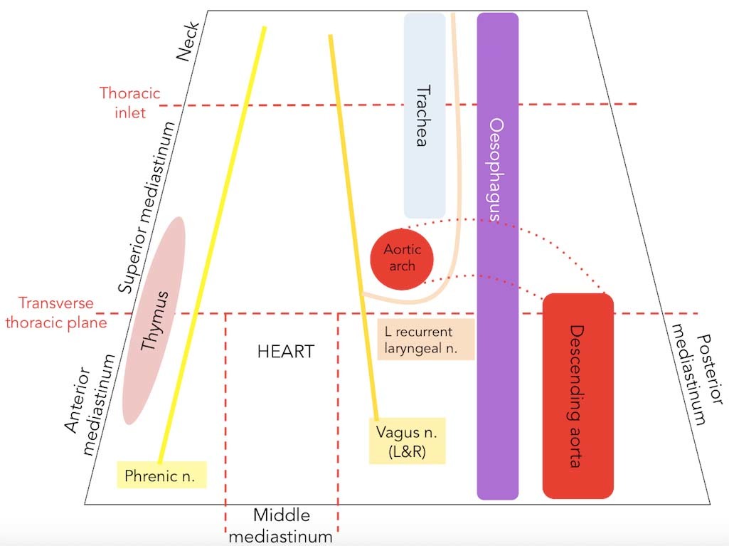
Divisions
mediastinum is the central compartment of the chest and is divided into superior and inferior parts inferior part is the larger of the two and is further subdivided into the anterior inferior, the middle inferior and the posterior inferior mediastinum. thoracic plane (of Ludwig), which is an artificial plane T4-5 to sternal angle
Contents
The mediastinum contains the heart and its major vessels, the trachea, the oesophagus, the remnant of the thymus (in adults), the thoracic duct and the phrenic nerves and cardiac nerves, including the vagus. It also contains the lymph nodes that can become massively enlarged in diseases such as lymphomas.
Hilar enlargement
Massive hilar lymphadenopathy is characteristic of lymphoma, but hilar and paratracheal lymph node enlargement is also typical of sarcoidosis. Enlargement may be due to metastatic disease and also to pulmonary hypertension
Abnormalities of the size, shape and contours of the myocardium
Numerous conditions can alter the cardiac shadow. These include cardiomegaly due to left and right ventricular hypertrophy or dilated and obstructed cardiomyopathy; left atrial enlargement, secondary, for example, to mitral stenosis, which may manifest as a double right heart border; cardiac outline is also changed by pericardial effusion. The definition of the heart borders may be lost; if on the left it may be due to consolidation of the lingula, if on the right it may be due to consolidation of the middle lobe of the lung.
Abnormalities of the vessels
Abnormalities of the vessels. This applies particularly to the thoracic aorta, which may be distorted by aneurysmal dilatation and increase the size of the aortic knuckle while also displacing the trachea.
Mediastinal widening.
Mediastinal widening. This can be a common artefact, but otherwise can be due to mediastinal masses, particularly paratracheal nodes in the superior mediastinum, and to vascular abnormality.
Pneumomediastinum.
Pneumomediastinum. This may follow penetrating trauma or perforation of the trachea or oesophagus. It may also occur in patients with chronic lung disease. If the source of the air is pulmonary, then it tracks from ruptured alveoli along the vascular sheaths which accompany the bronchi, and once at the hilum extends proximally into the mediastinum. If air in the mediastinum is under tension it can occasionally compromise cardiac output, but in itself it is usually innocuous and self-limiting. This may not be true of the precipitating cause.
masses
Other masses. These include retrosternal goiters and lesions of the thymus
Mediastinoscopy
Mediastinoscopy. This allows the biopsy of mediastinal masses either for diagnosis or for the staging of disease. The commonest approach is via a relatively small incision in the suprasternal notch. The difficulties faced by the anaesthetist will be influenced largely by the varied nature of the disease. Tracheal compression, for example, may make airway management problematic. Lung cancer is likely to be accompanied by chronic obstructive pulmonary disease and ischaemic heart disease. A thymic mass may be associated with myasthenia gravis.
Potential complications
include massive haemorrhage, pneumothorax, air embolism and tracheobronchial injury. However, in many cases mediastinoscopy has been superseded by endobronchial ultrasound-guided fine needle aspiration (EBUS), which is a considerably less invasive technique that spares patients most of the complications outlined here.