Maternal Changes in Pregnancy
暂无描述。系统推荐的高质量记忆内容,适合每天坚持背诵学习。
卡片预览 (24 张)
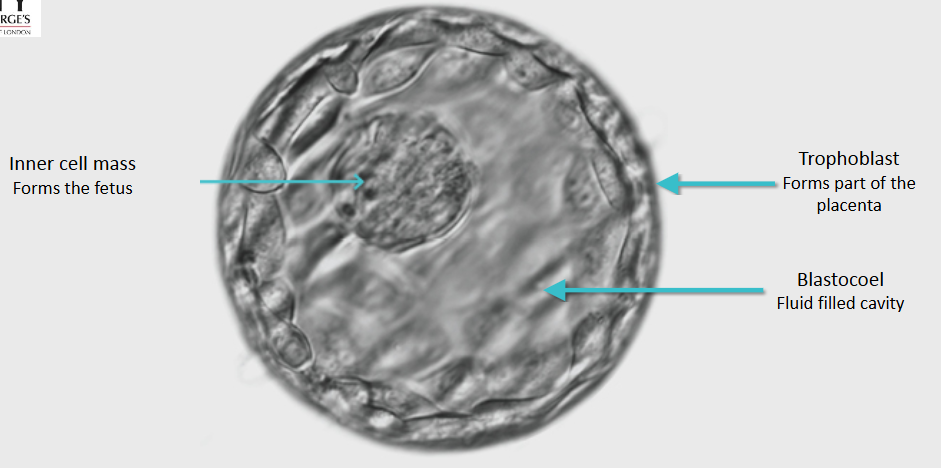
Descibe the human blastocyst?
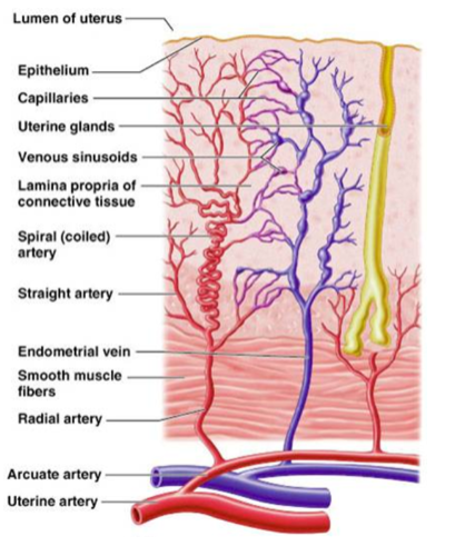
What are the key endometrial changes during the implantation window and early pregnancy?
• Maximum endometrial changes: ~7 days after ovulation. • Implantation window: 6–10 days after LH spike. • Pre-decidualization: 9–10 days after ovulation; decidual cells begin covering uterine surface. • Decidualization in pregnancy: Cells become modified with lipids and glycogen; form the maternal part of the placenta. • Glandular secretions contain growth factors, adhesion molecules, nutrients, vitamins, matrix proteins, and hormones to support implantation.
What roles do decidual cells, syncytiotrophoblast, and chorionic gonadotropin play in early pregnancy?
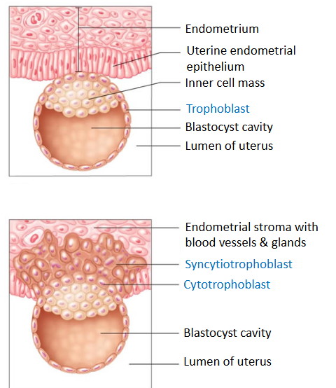
• Decidual cells: On the endometrial surface, fill with lipids and glycogen → become the maternal part of the placenta. • Syncytiotrophoblast: Forms by cell fusion, creating a multi-nucleated cytoplasmic mass that invades the endometrium. • Chorionic gonadotropin (hCG): Acts as an autocrine growth factor for the blastocyst
What are the key stages of blastocyst implantation in the first 16 days?
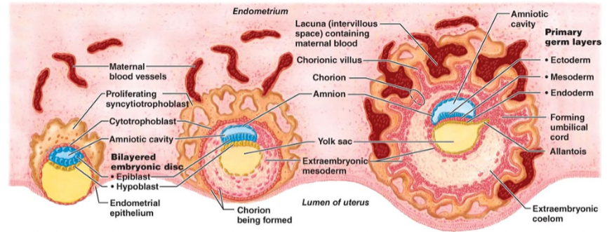
Day 7–8: * Syncytiotrophoblast erodes the endometrium. * Embryonic disc forms epiblast (future amnion) and hypoblast. * Epiblast develops the fluid-filled amniotic cavity. Day 12: * Implantation complete. * Extraembryonic mesoderm forms beneath the cytotrophoblast. Day 16: * Cytotrophoblast + associated mesoderm = chorion. * Chorionic villi extend into maternal tissue. * Lacunae filled with maternal blood mingle with villi, establishing early maternal-fetal circulation.
Describe the maternal-fetal interface?
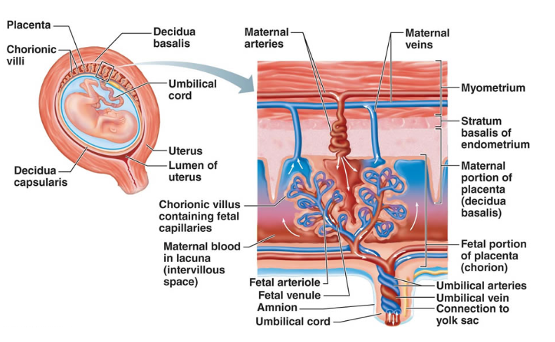
• The maternal-fetal interface is a specialized, dynamic zone of interaction between maternal uterine tissues (decidua) and fetal-derived placental tissues (trophoblasts). • It functions as a complex immunological, physiological, and physical barrier, enabling nutrient/gas exchange and fostering tolerance toward the foreign fetus while protecting against infections.
What is the role of human chorionic gonadotrophin (hCG) in early pregnancy?
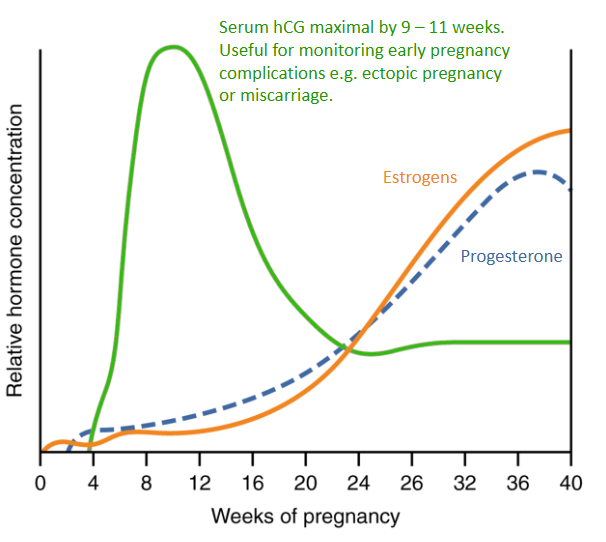
Source: Secreted by the syncytiotrophoblast; rapid rise is the basis of pregnancy tests. Function: * Prevents death of the corpus luteum, so the endometrium is maintained. * Corpus luteum continues producing estrogen and progesterone. Effect: Maternal systems undergo rapid changes in response to luteal and later placental steroids.
What are the key roles of placental steroids in pregnancy and their synthesis pathways?
Synthesis: * Derived from fetal and maternal adrenal steroids. * Placenta lacks 17α-hydroxylase and 17,20-lyase, so some conversions require maternal/fetal contribution. Progesterone: Synthesized directly from cholesterol. Functions: * Decidualization (corpus luteum support) * Smooth muscle relaxation → uterine quiescence * Mineralocorticoid effect → cardiovascular adaptations * Breast development (glands & stroma) Estrogens (Estradiol E2, Estriol E3): Synthesized via aromatase. Functions: * Uterine hypertrophy * Cardiovascular changes * Increased clotting factor production → haemostasis * Breast development * Metabolic changes (insulin resistance)
What are the patterns and components of maternal weight gain during pregnancy?
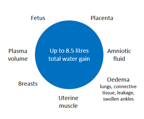
Average total gain: 9–13 kg. Components: * Fetus & placenta: ~5 kg * Fat & protein: ~4.5 kg * Body water: ~1.5 kg (intravascular, interstitial, intracellular) * Breasts: ~1 kg * Uterus: 0.5–1 kg Timeline: * First 20 weeks: ~2 kg total * After 20 weeks: ~0.5 kg per week until 40 weeks Clinical notes: * Failure to gain or sudden weight change requires investigation. * Constant monitoring can cause anxiety.
How does basal metabolic rate (BMR) change in pregnancy, and what are the energy and glucose implications?
BMR increase: * Mid-gestation: +350 kcal/day * Late gestation: +250 kcal/day * Energy use: ~75% for fetus & uterus, 25% for maternal respiration Energy storage: 9 kcal ≈ 1 g fat → 350 kcal ≈ 40 g fat Glucose: Maternal glucose rises to cross the placenta for fetal needs
How does maternal and fetal glucose metabolism change across pregnancy?
First trimester Maternal reserves * Pancreatic cells increase in number raising circulating insulin so more glucose is taken up into tissues. Fasting serum glucose decreases. Second trimester Fetal reserves * Placental Lactogen causes insulin resistance, ie less glucose into stores and increase in serum glucose. Transfer of glucose to fetus. * Increased glucose level in blood during 2nd trimester. Glucose is transported across placenta as fetal energy source. Fetus stores some in liver.
What is gestational diabetes?
• Defined as transient glucose intolerance, which gets corrected after the end of the pregnancy • Occurs due to a predominance of anti-insulin hormones • Pregnancy leads to fasting hypo-glycaemia and post-prandial hyper-glycaemia • Linked with obesity and ethnicity • Can be controlled mostly by dietary adjustment
How is gestational diabetes screened and diagnosed?
• Screening test: 75 g oral glucose tolerance test (OGTT). • Timing: 26–28 weeks of pregnancy, after an overnight fast. • Urine testing: Unreliable due to reduced renal threshold. • Blood glucose: Fasting and 2-hour post-glucose measured. • WHO thresholds: • Fasting: ≥ 5.6 mmol/L • 2-hour: ≥ 7.8 mmol/L
What are the maternal and fetal effects of gestational diabetes?
Maternal Effects: * Polyhydramnios (excess amniotic fluid) * Increased risk of infections * Preterm birth * Instrumental delivery (forceps or vacuum) * Later development of diabetes Fetal/Neonatal Effects: * Macrosomia (large baby) * Shoulder dystocia * Neonatal hypoglycaemia * Jaundice * Congenital abnormalities: not increased * Perinatal morbidity * Stillbirth
What are the long-term risks and treatment considerations for gestational diabetes?
• Long-term risk: Higher chance of developing Type II diabetes later in life (acts like a stress test). • Treatment: • Mostly dietary management. • Sometimes requires Metformin or insulin for glucose control. • Drugs that stimulate insulin production are contraindicated in pregnancy. • Classification: Women needing insulin during pregnancy should be re-classified as Type II diabetes complicating pregnancy.
How do estrogen, progesterone, and pregnancy hormones affect maternal fluid balance and vascular system?
• Sodium & Blood Volume: • High estrogen and progesterone act like mineralocorticoids → retain sodium → increase blood volume. • RAAS Activation: • Placental renin + estrogen ↑ angiotensinogen → ↑ angiotensin II and aldosterone. • Despite high ANG II, vascular resistance decreases because progesterone reduces vasoconstriction sensitivity. • Connective tissue: Ligaments absorb water → become softer. • Fluid/osmoregulation: • Reset osmostat → decreased thirst threshold. • Oncotic pressure decreases (lower albumin).
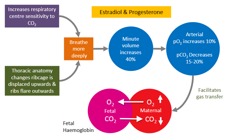
How does oxygen consumption change during pregnancy?
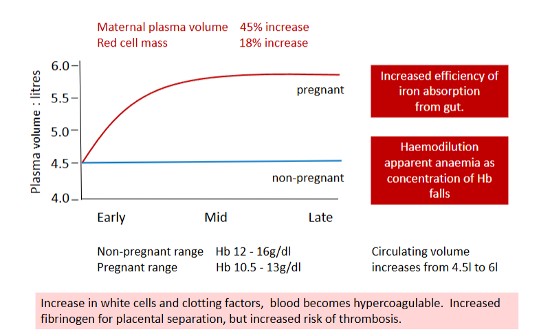
How does maternal blood change during pregnancy?
What are the key cardiovascular changes during pregnancy?
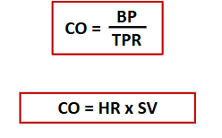
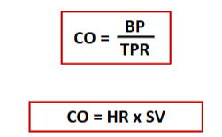
• Mechanical changes: • Expanding uterus pushes the heart → alters ECG and heart sounds • Peripheral circulation: • Peripheral vasodilation (via nitric oxide, ↑ by estrogen/E2) • ↓ Total peripheral resistance (TPR) by 20–30% • Cardiac output (CO): • Increases (up to ~40% by 28 weeks) • Begins as early as 3 weeks • Due mainly to ↑ stroke volume + slight ↑ heart rate (8–10 bpm) • Blood pressure: • Decreases in 1st and 2nd trimesters • Clinical significance: • Increased cardiac workload can worsen pre-existing conditions (e.g., aortic valve disease, pulmonary hypertension)
What are the key vascular (blood vessel) changes in the cardiovascular system during pregnancy?
• Increased cardiac output + steroid-induced vasodilation Reduced peripheral resistance → increased blood flow to: * Uterus * Placenta * Muscle * Kidneys * Skin Neo-angiogenesis: * Formation of new blood vessels (via VEGF/PLGF) * Extra skin capillaries → spider naevi and improved heat loss Overall state: * Low-pressure, high-volume circulation during pregnancy
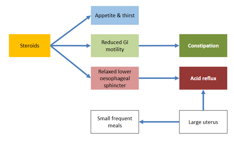
Describe the changes in the gastrointestinal tract during pregnancy?
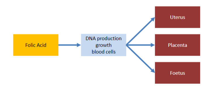
Explain the importance of folic acid - dietry supplement?
• Supplementation advised up to 400μg/day until week 12 • Ideally 3 months before pregnancy. • Deficiency can lead to birth defects eg spina bifida (neural tube defects).
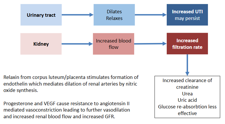
Describe the changes in the urinary system during pregnancy?
• Relaxin from corpus luteum/placenta stimulates formation of endothelin which mediates dilation of renal arteries by nitric oxide synthesis. • Progesterone and VEGF cause resistance to angiotensin II mediated vasoconstriction leading to further vasodilation and increased renal blood flow and increased GFR
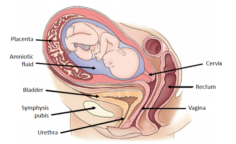
Describe urinary frequency
Frequent urination is a common, often daily, symptom of pregnancy caused by hormonal shifts (hCG, progesterone), increased blood volume, and pressure from the growing uterus on the bladder.
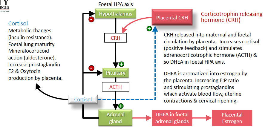
How does placental CRH regulate the fetal–maternal endocrine system and contribute to labour?
Placental CRH: * Released into maternal and fetal circulation * Stimulates ACTH → ↑ cortisol (positive feedback loop) * Activates fetal HPA axis → ↑ DHEA production Cortisol effects: * Insulin resistance (metabolic changes) * Promotes fetal lung maturity * Has mineralocorticoid effects (via aldosterone) DHEA pathway: * Fetal DHEA → converted (aromatized) in placenta → estrogen Estrogen effects: * ↑ Estrogen:Progesterone ratio * ↑ Prostaglandin (PGE2) and oxytocin production Outcome: * Increased uterine blood flow * Uterine contractions * Cervical ripening → contributes to onset of labour