Haematology Pictures
No description yet. This curated deck is ready for daily study.
Card preview (12)
What type of anaemia does this smear show? When would this be seen?
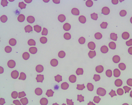
Normochomic normocytic (erythrocytes all similar size) Seen with mild non-regenerative amaemia
What type of anaemia does this smear show? When would this be seen?
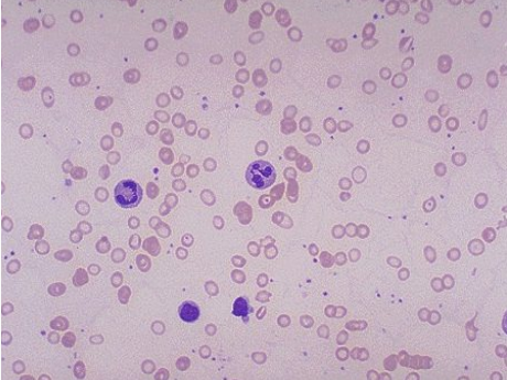
Microcytic, seen in Fe deficiency, PSS, hepatic failure (iron deficiency allows one more division of red cells as Hb concentration determines when division stops) • may be seen in Akitas (congenital)
What type of anaemia does this blood smear show? What breed may be predisposed to this?
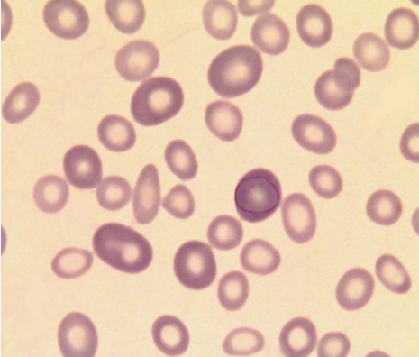
Macrocytic regenerative - polychromatophils can be seen, larger than RBCs • Poodles
How would these cells be described? When are these kind of RBCs seen?
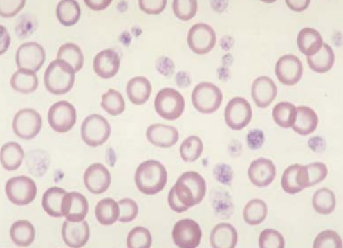
Hypochromic cells, relating to Hbg conc • Fe insufficiency or incorporation -> microcytosis (cells are thinner hence appear paler, not necessarily a decrease in diameter)
What type of cell can be seen here? What does this indicate?
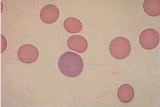
Polychromatophil - indicates regeration as it is an immature RBC in the circulation. Will continue to mature in blood stream so may be pale blue. Have this appearance with Diff-Quik or Giemsa stains.
What cells can be seen here? What are these the same as?
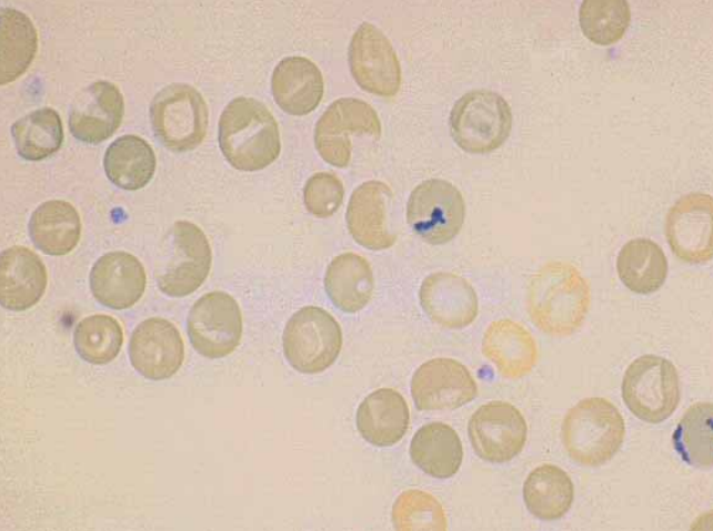
Reticulocytes - same as polychromatophils but stained with new methylene blue to precipitate out the RNA
What signs of regeneration can be seen on these smears?
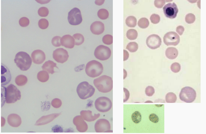
Polychromasia, anisocytosis, macrocytosis, nRBCs, Howell-Jolly bodies [top right], codocytoisis, basophilic stippling [Bottom right]
What type of abnormal cell can be seen here? Ehat does this indicate?
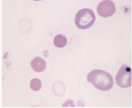
Ghost cell - not sure what this shows!
What abnormal RBC morphology can be seen here?
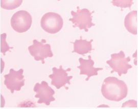
Shear products (keratocytes, shistocytes, acanthocytes)
What can be seen here? What does this indicate?
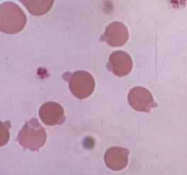
Heinz bodies, indicate oxidative damage
What are these cells? What do they indicate?
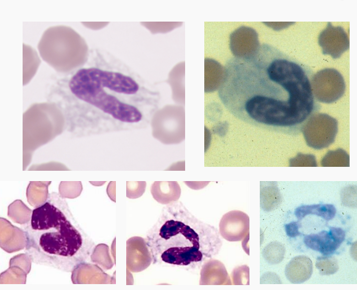
Band neutrophils - indicate left shift (Acute infection)
What has happened here?
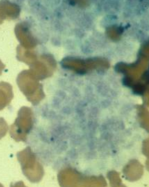
Platelet clumping