3. Massive Haemorrhage: Compensatory Responses and Management
No description yet. This curated deck is ready for daily study.
Card preview (10)
The normal compensatory responses to the loss of intravascular volume.
function of the circulation is to distribute the cardiac output to tissues sufficient to meet their metabolic demands. Any progressive loss of circulating volume is accompanied by a • redistribution of flow aimed to ensure that the brain and myocardium continue to receive oxygenated blood. As blood loss continues, the decreases in venous return, right atrial pressure and cardiac output activate baroreceptor reflexes (mediated by stretch-sensitive receptors in the carotid sinus and aortic arch). This is an immediate response The decreased afferent input to the medullary cardiovascular centres inhibits parasympathetic and enhances sympathetic activity
Sympathetic increase
There follows an increase in cardiac output together with alterations in the resistance of vascular beds in an attempt to maintain tissue perfusion. These changes are mediated via direct sympathetic innervation, by circulating humoral vasopressors such as adrenaline, angiotensin, noradrenaline and vasopressin, and by local tissue mediators, including hydrogen ions, potassium, adenosine and nitric oxide. Hypovolaemia encourages movement of fluid into capillaries, the decreased capillary hydrostatic pressure favouring absorption of interstitial fluid with a resultant increase in plasma volume and restoration of arterial pressure towards normal (Starling forces).
The hypothalamo–pituitary–adrena
The hypothalamo–pituitary–adrenal response is also important, although it is slower. Reduced renal blood flow stimulates intrarenal baroreceptors which mediate renin release from the juxta-glomerular apparatus. Renin converts circulating angiotensinogen to angiotensin I, from which angiotensin II (AT II) is formed in the lung. AT II is a potent arteriolar vasoconstrictor that stimulates aldosterone release from the adrenal cortex and arginine vasopressin (ADH) release from the posterior pituitary. ADH release is also stimulated by atrial receptors, which respond to the decrease in extracellular volume. These changes enhance sodium and water reabsorption at the distal renal tubule as the body attempts to conserve fluid. Sympathetic stimulation also mediates secretion of catecholamines and cortisol.
Inflammatory response
significant inflammatory response to major haemorrhage. Hypoperfused and hypoxic endothelium releases the familiar inflammatory mediators, oxidants and cytokines as is seen in sepsis and reperfusion injuries, and these may similarly mediate coagulopathy and a systemic inflammatory response syndrome (SIRS). SIRS). This phenomenon emphasizes the importance of flow rather than pressure. Endogenous catecholamines and exogenous vasopressors may increase arterial pressure, but at the level of the microcirculation, pre-capillary arteriolar constriction will simply decrease flow through the capillary bed, compromise tissue perfusion further and accelerate the inflammatory response
Acute Trauma Coagulopathy (ACT).
Acute Trauma Coagulopathy (ACT). Major trauma is associated with the development of coagulopathy that is proportional to the severity of the injury and which is now specifically described as ‘acute trauma coagulopathy’. Its mechanisms have not fully been elucidated, but it is likely that critically underperfused vascular endothelium accelerates activation of the coagulation cycle with consumption particularly of Factor V and fibrinogen (Factor I). The effectiveness of coagulation is also compromised by hypothermia and acidaemia, both of which may frequently complicate severe trauma.
The Metabolic Acidosis Associated with Blood Loss
Lactic acidosis: decreased tissue perfusion causes a progressive decline in aerobic metabolism, which is accompanied by a compensatory increase in anaerobic metabolism. This shift to anaerobic metabolism results in a decrease in energy production and the development of a metabolic acidosis In the absence of molecular oxygen, the final acceptor is missing and so NADH accumulates. The lack of NAD+ effectively blocks the TCA cycle, and so pyruvate (CH3-C = O-COOH) also accumulates (at the ‘entrance’ to the cycle). NADH and pyruvate react to form lactate (CH3-HCOH-COOH) and NAD The lactate then diffuses out of the cell to accumulate as lactic acid; NAD+ meanwhile allows anaerobic glycolysis to proceed.
Summary of clinical features
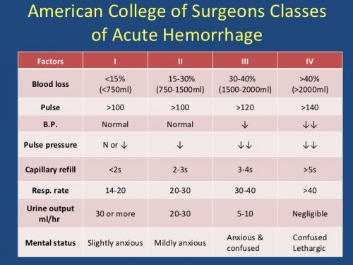
: redistribution of blood flow is responsible for the typical pallor, cold peripheries, peripheral cyanosis and oliguria. Sympathetic stimulation explains the tachycardia and the increase in respiratory rate. Carotid chemoreceptors also stimulate ventilation in response to changes in PaO2, PaCO2 and pH. Systolic blood pressure is a relatively crude index which may show little change until substantial volumes have been lost, particularly in young patients. The pulse pressure may be more useful; as blood loss continues, it narrows, and the mean arterial pressure may increase. This occurs because diastolic blood pressure is under the influence of catecholamines which rise in response to haemorrhage. Capillary refill time is a simple and effective measure. A delay of more than 2 seconds is abnormal, and trends can be used to gauge the effectiveness of fluid resuscitation. Confusion or other changes in mental state indicate cerebral hypoxaemia and hypoperfusion.
Massive Haemorrhage: Principles of Management
The management of the patient with massive haemorrhage continues to evolve, particularly in the light of military trauma management. Airway and Breathing remain important components of the ABCDE assessment and management mantra, but C for Circulation is now less obvious. Controlling the bleeding at source is the key priority, whether it be due to trauma, post-partum haemorrhage or a ruptured aortic aneurysm. In some situations, particularly following trauma, this will be damage-control resuscitation prior to definitive surgical repair.
Resus goals
adequate tissue perfusion, and although from the anaesthetic perspective it may seem counter-intuitive, it can be deleterious to resuscitate the patient to euvolaemia and normotension. This may contribute a dilutional element to the coagulopathy, and may reduce the oxygen-carrying capacity of the circulation while jeopardizing flow through the capillary bed, as described earlier Current practice is to aim for ‘permissive hypotension’ with a systolic blood pressure of around 80 mmHg and the more cautious titration of fluids against response. In the acute phase, moreover, it is clear that the optimal resuscitation fluids are blood, coagulation factors (in fresh frozen plasma and cryoprecipitate) and platelets. Military studies support initial transfusion of fresh frozen plasma (FFP) and packed red blood cells (PRC) in a 1:1 ratio, with platelets and cryoprecipitate given ideally in response to point of care coagulation tests, with tranexamic acid given as routine. Civilian protocols tend to vary, with FFP and PRC sometimes given in a 1:2 ratio. Although it may seem formulaic, it is clearly associated with better outcomes than the previous practice of transfusing red cells and then giving coagulation factors after a sometimes lengthy wait for a clotting profile, which in a dynamic situation may be inaccurate by the time that that is measured. (For general complications of blood transfusion, including those associated with rapid infusion, see under ‘Complications of Blood Transfusion’.)
blood loss parameters