Sub-cellular Organelles
No description yet. This curated deck is ready for daily study.
Card preview (12)
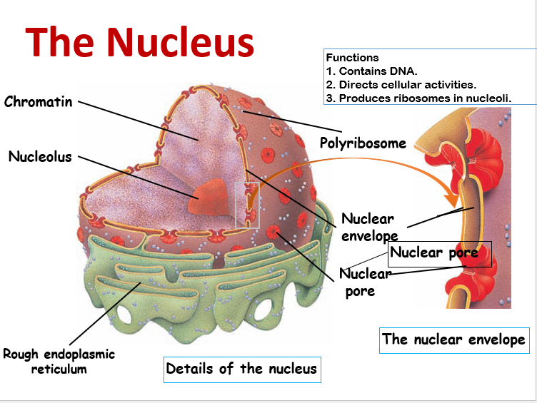
The Nucleus
Control centre of cell. Cells generally have a single nucleus: mature RBCs have none, unlike skeletal muscle cells that have multiple nuclei. contains genes – determine cell characteristics: determine types of proteins, promote and control reproduction: gene cloning. split by mitosis with equal share of DNA. chromatin – DNA, RNA and proteins: DNA packaging, structural reinforcement and protect against damage. Nuclear envelope – 2 membranes: outer - in continuity with E.R. peri-nuclear membrane – inner layer; has nuclear pores. pores consists of a circular arrangement of proteins surrounding a large central opening. control movement of proteins and RNA across envelope. Nucleoplasm – suspending media; polymerase enzymes. Nucleolus – site of ribosome synthesis and assembly
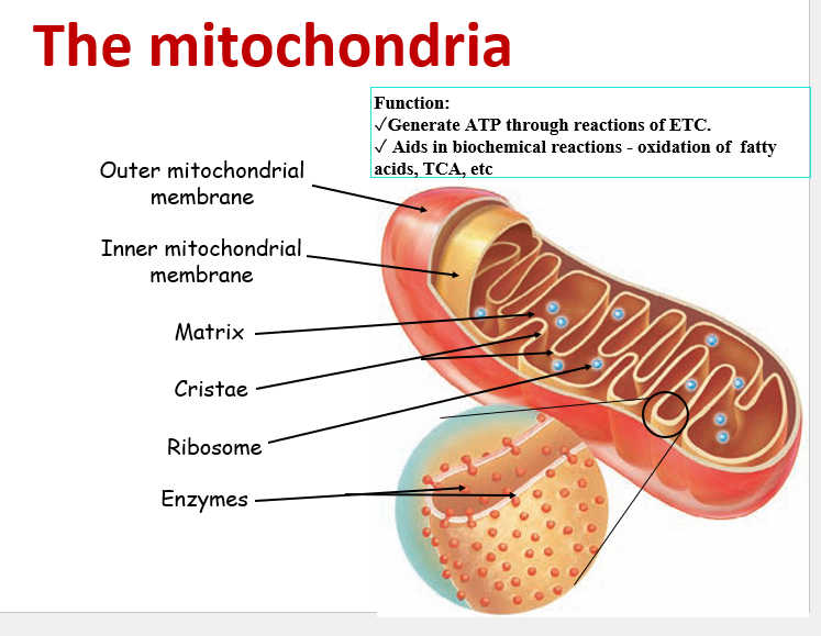
The Mitochondria
Varying size & shape - elongated or rod shaped. Powerhouse of Cell: range from a few as 100 or as many as several 1000 depending on the activity of the cell. Throughout cytoplasm. Outer and inner membranes. Outer membrane – continuous lipid in nature. freely permeable (allows small molecules) protein pores Inner – mainly protein in nature. selectively permeable. folds to form cristae.( ↑ surface area ) cardiolipin (a unique, dimeric phospholipid crucial for mitochondrial function, especially energy production (ATP synthesis) and membrane structure, acting as a scaffold for respiratory proteins). oxidative phosphorylation machinery - ETC, ATP synthase Inter-membrane space – adenylate kinase: phosphotransferase (AMP, ADP and ATP) Several isoenzymes - substrate, H+ donor, tissue distribution and subcellular localization. ATP+AMP⇔ 2ADP Matrix- has specific circular DNA , ribosomes and enzymes. Mitochondrial DNA – maternal inheritance. Functions: site of energy production site of metabolic pathways.- TCA cycle, Urea Cycle, F.A Oxidation etc.
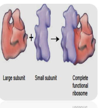
Ribosome Subunits
Sites of protein synthesis. high content of r-RNA. they are present free in cytoplasm or attached to RER. consists of two subunits. large & small subunits Prokaryotes: 70S = 50S +30S Eukaryotes: 80S = 60S + 40S (S) Svedberg unit: mass shape & size
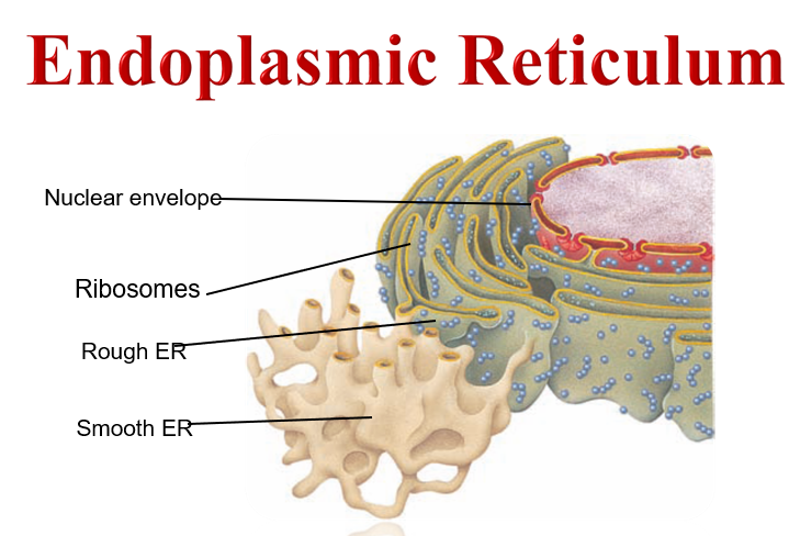
Endoplasmic Reticulum
Interconnected network of tubules and vesicles – endomembrane system. Flattened membranous structures – cisternae. stacks of about 3-7. RER extends from nucleus to plasma membrane. Two types of ER : rough and smooth ER. RER- synthesize, sort proteins; lipid mod. SER- synthesize phospholipids, steroids + cholesterol in many tissues. SER - site of Glycogen metabolism. removes the phosphate group from G-6-P; release free glucose in blood. important in homeostatic regulation of blood glucose In liver & Kidney - detoxifies drugs, toxins & carcinogens. Sarcoplasmic reticulum - stores & releases calcium ions: specialized ER surrounds each myofibril. regulates cytoplasmic calcium levels in muscle cells.
Golgi Apparatus
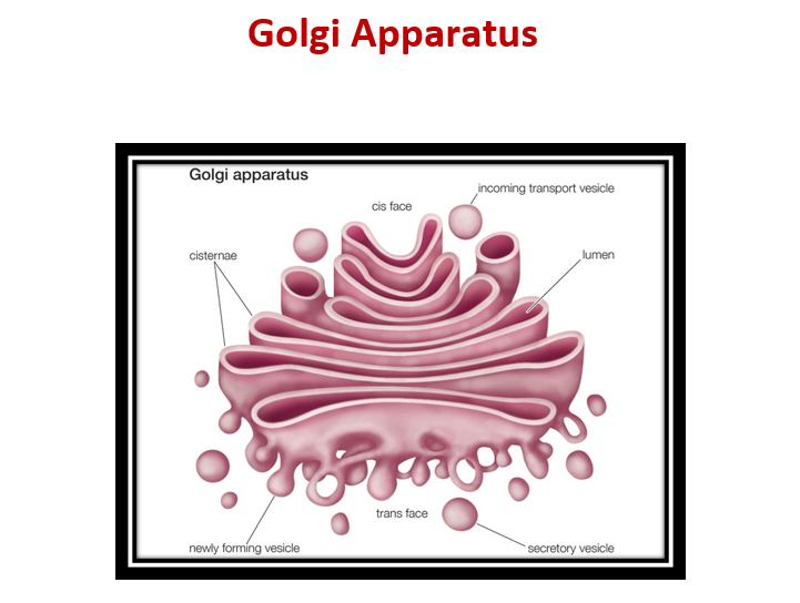
Consists of 3 – 20 flattened membranous sacs – cisternae: also part of endo-membrane. Cis face towards ER: vesicles from ER fuse releasing contents. contents modified in lumen for transport. Trans face distal to ER: Proteins not intended for ER or Golgi travel through cisternae network. trans Golgi network – further protein modification. Hence, fundamental in vesicular transport. Trans-golgi Network - cis, medial and trans Folding + sorting Vesicular transport Prominent in protein releasing cells: sorts, packs, modifies and transport protein from RER. proteins packaged into membrane bound vesicles. process proteins for secretion: protects integrity adhesion proteins - interns cell-cell adhesion
Lysosome
Tiny membrane-enclosed vesicles from Trans-Golgi. Suicidal bags – Hydrolase: digest protein, nucleic acid, lipids and sugars 60 kinds of powerful digestive and hydrolytic enzymes: optimum pH 5 – more acidic than cytoplasm. Helps in fertilization acrosome special lysosome – protease, phosphatase. Role in autophagy – prog. cell ‘eating’: rid cell of old organelles. isolates targeted structures and forms autophagosome. autophagosome fuses with lysosomal bodies – release contents. Role in phagocytosis by W.B.C. - PMN chemotaxis (Polymorphonuclear Leukocyte/Neutrophil) : Lysosomal storage diseases – Genetic diseases, due to absent / deficient lysosomal enzymes. e.g., Niemann pick disease, (a group of rare, inherited metabolic disorders where fats (lipids) build up in cells, damaging organs like the brain, liver, spleen, and lungs) Gaucher’s disease ( a rare, inherited lysosomal storage disorder caused by a genetic mutation (in the GBA gene) leading to a deficiency of the glucocerebrosidase enzyme, resulting in fatty substances (glucocerebroside) building up in cells, particularly macrophages, in the liver, spleen, bone marrow) Tay-Sachs disease: which causes mental retardation and blindness, is caused by the loss of hexosaminidase A, a lysosomal enzyme that catalyzes the biodegradation of gangliosides (fatty acid derivatives).
Peroxisome
Cytoplasm
Cytoskeleton
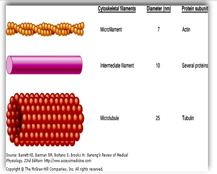
A network of protein filaments that extends throughout the cytoplasm. Anchored to plasma membrane. Three types of filamentous proteins: microtubules intermediate filaments microfilaments Provides shape to cell - internal framework. Helps in uptake of materials into cell – ‘track’ network for sub-cellular cargo. Helps in internal movement of cell organelles, movement of cells and muscle contraction. Helps in Cell division.
Microfilament
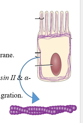
Thinnest elements of the cytoskeleton. Composed of the protein actin: β- and γ-actin. Form a meshwork under plasma membrane. Stress fibres: non-muscle cells – non-muscle myosin II & α-actin. provide force for cell adhesion + migration. Other functions : mechanical support - strength + shapes of cells: e.g. - microvilli are rich in microfilaments. involved in muscle contraction and cell division.
Intermediate Filament
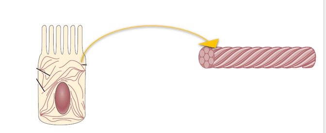
Polymers of long rod like proteins. These filaments are thicker than microfilaments but thinner than microtubules. Include keratin, desmin, neurofilaments etc. Functions: provide mechanical support to the cell. help in intercellular attachment. provide strength and rigidity to neurons. major structural role in skin and hair cells structure to mesenchymal cells - vimentin Nuclear support - lamin
Microtubules
Long, Hollow, unbranched, polar cylinders. made up of protein “tubulin” α- + β-tubulin The largest of cytoskeletal components. Major components of axons + dendrites. Functions: microtubules help in structural support and maintaining cell shape. helps in movement of organelles, secretory vesicles and exocytosis. formation and function of mitotic spindle. movement of cilia and flagella. Disorder: Primary ciliary dyskinesia- refers to a set of inherited disorders that limit ciliary structure and/or function. clinical effect- recurrent upper & lower respiratory tract infection to slow the mucociliary escalator and result in airway obstruction and increased infection Male infertility - Dysregulation of ciliary function in sperm cells has also been well characterized to result in loss of motility.