Theme 1: Signalling in the Cardiac Myocyte
No description yet. This curated deck is ready for daily study.
Card preview (24)
Is cardiac muscle striated?
Yes
Describe the nuclei of cardiac muscle.
Each cell only has one nucleus, which is centrally located.
What is unusual about the shape of cardiac myocytes?
They are branched.
What joins cardiac muscle cells?
Intercalated discs
What is the distinguishing characteristic of cardiac muscle in histology?
The presence of dark transverse lines between cells -> These are intercalated discs.
What are intercalated discs?
Structures that interface between adjacent cardiac muscle cells and support synchronised contraction.
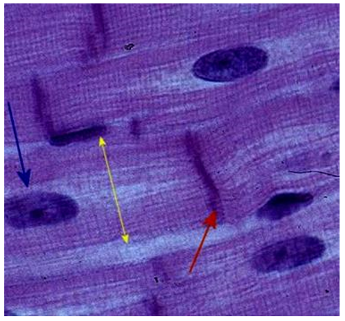
What are the 3 components of intercalated discs?
• Desmosomes • Adherens junctions • Gap junctions
What is the function of desmosomes in intercalated discs between cardiac myocytes?
Bind cells together.
What is the function of adherens junctions in intercalated discs between cardiac myocytes?
Act as anchoring sites for actin filaments.
What is the function of gap junctions in intercalated discs between cardiac myocytes?
Provide continuity between adjacent cells and allow ions to pass between cells.
What is another name for adherens junctions in intercalated discs?
Fascia adherens (a.k.a. hemi Z-bands)
What is another name for desmosomes in intercalated discs?
Macula adherens
Summarise simply the structure and functioning of cardiac myocytes.
Branching mesh of mononuclear striated cells joined and electrically coupled by intercalated discs (desmosomes and gap junctions: electrically a ‘functional syncytium’).
Draw the structure of a sarcomere. Include the names of the different zones.
• A band (dark) -> All of the length of myosin filaments • I band (light) -> Just actin filaments • Z line -> Where actin attaches • H band -> Just myosin filaments • M line -> What myosin attaches to
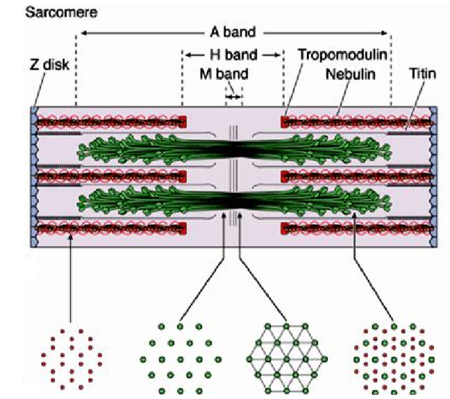
Label this section of cardiac muscle.
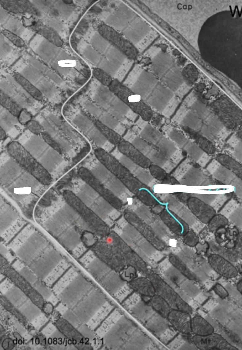
• Mt = Mitochondria • ln D = Border of the myocyte
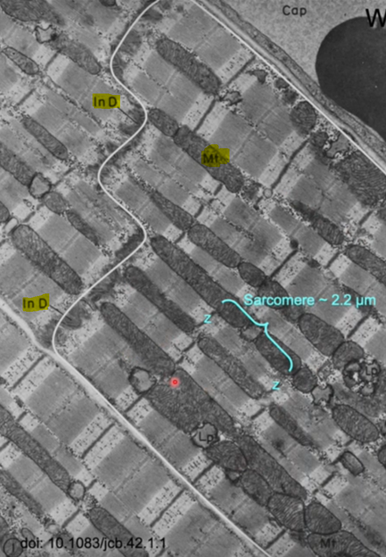
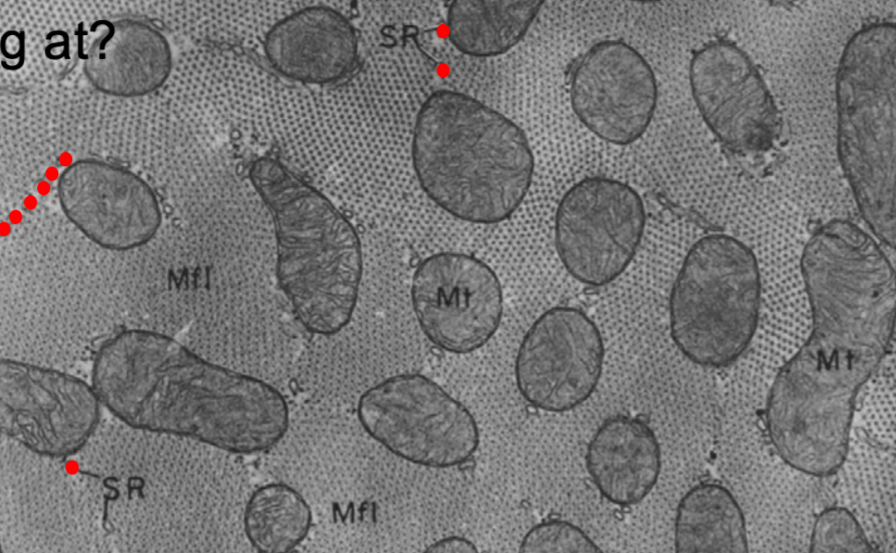
How is the sarcoplasmic reticulum arranged in cardiac muscle?
The SR run along myofibrils.
What are T tubules and cisternae?
• T tubules -> Invaginations of the plasma membrane into the myocytes (contain LTCCs) • Cisternae -> Parts of the sarcoplasmic reticulum that are adjacent to the T tubules (contain RyR)
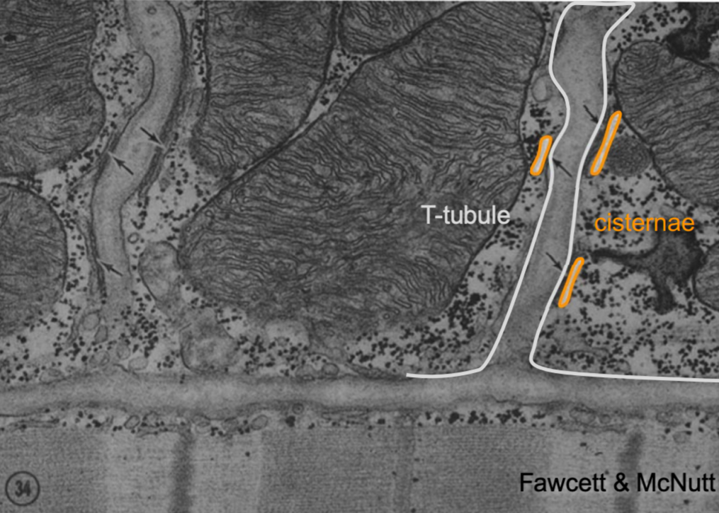
What are dyads?
• The dyad is a structure in the cardiac myocyte composed of a single T-tubule paired with a terminal cisterna of the SR. • It is like a triad in skeletal muscle.
At what point along the sarcomere in cardiac myocytes are the T-tubules?
At the z-lines (i.e. at the ends of the sarcomere)
Draw the subcellular organisation of cardiac muscle.
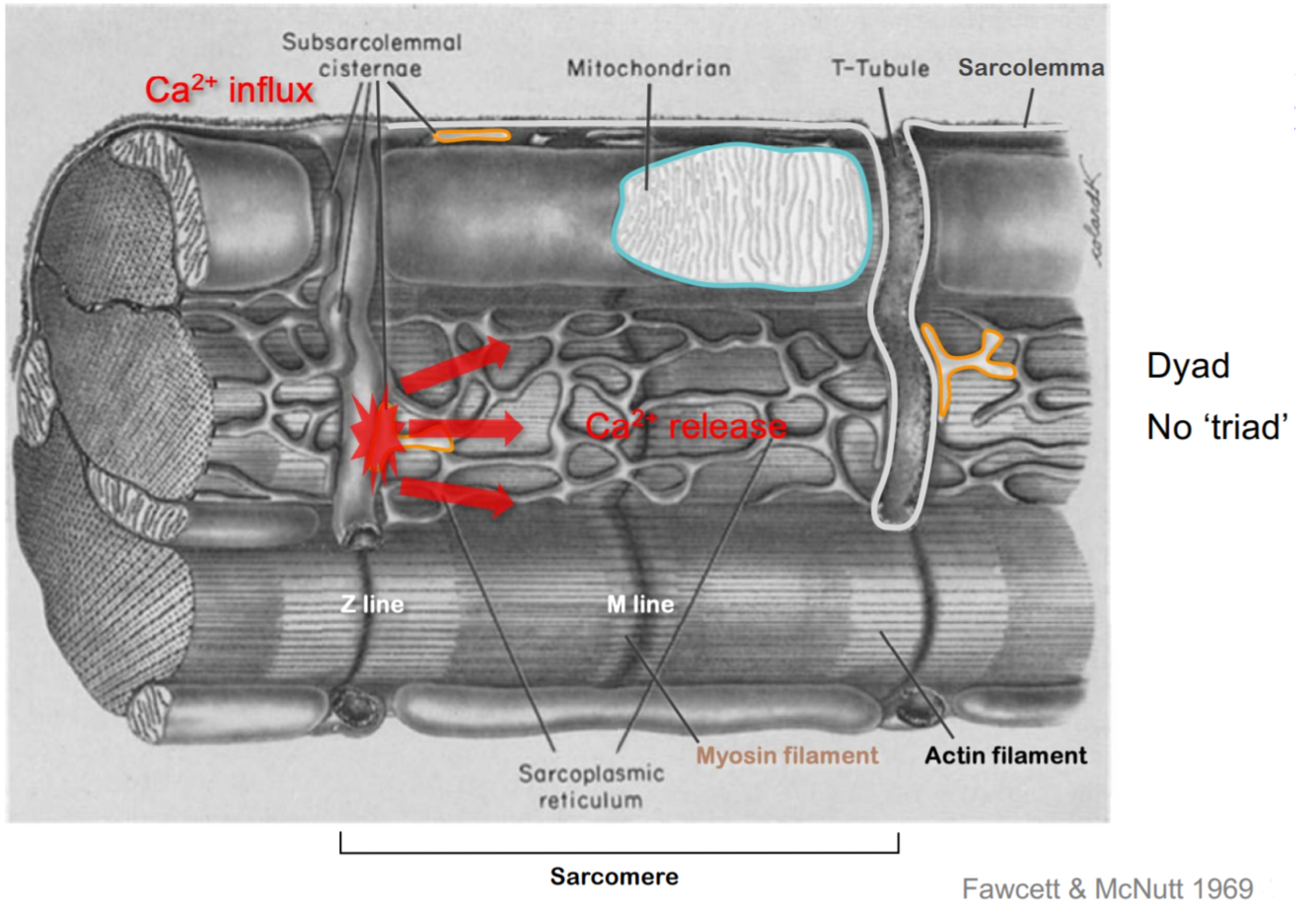
Describe excitation-contraction coupling in cardiac muscle.
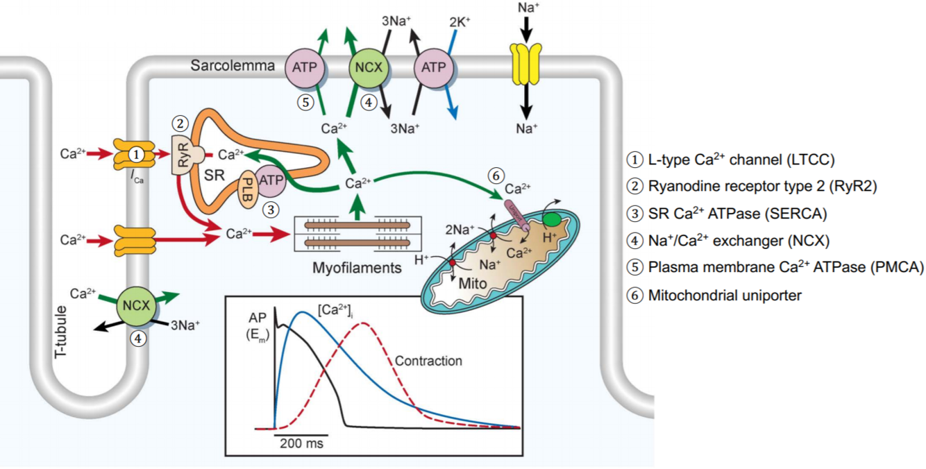
• Depolarisation of the sarcolemma travels along the membrane and into the T-tubule • T-tubules form dyads with cisternae of sarcoplasmic reticulum • Sarcolemma depolarisation causes opening of L-type Ca2+ channels (LTCC) in the sarcolemma • Calcium entry activates ryanodine receptors (RyR) on the sarcoplasmic reticulum -> This is called calcium-induced calcium release (CICR) • Calcium exits via the RyR into the cytosol • Calcium activates troponin C and triggers contraction
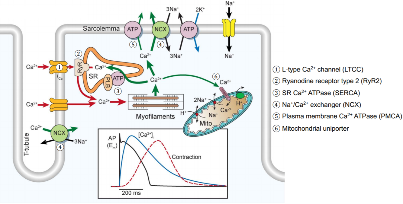
Draw the different calcium fluxes that occur within cardiac myocytes.
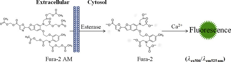
Give an example of a fluorescent calcium indicator and how it works.
Fura-2 acetoxymethyl ester (Fura-2 AM): • Extracellularly, it is not fluorescent and is in its ester form • This enables it to enter cells • Once it the cell, it is broken down by endogenous methylesterases, removing the acetoxymethyl groups • This leaves Fura-2, which is the indicator component that binds calcium and then gives off fluorescent light • The light can be detected by fluorescence microscopy or a fluorometer
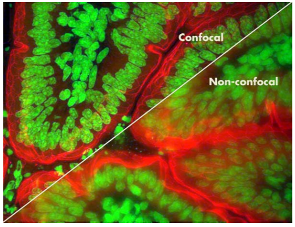
What microscopy technique can be used along with calcium-sensitive dyes to visualise calcium release in subcellular compartments?
Confocal fluorescence microscopy -> It essentially removes the out of focus parts of the image, leading to a clearer image