Chapter 1: The Science of Cognition
No description yet. This curated deck is ready for daily study.
Card preview (24)
Frontal lobe
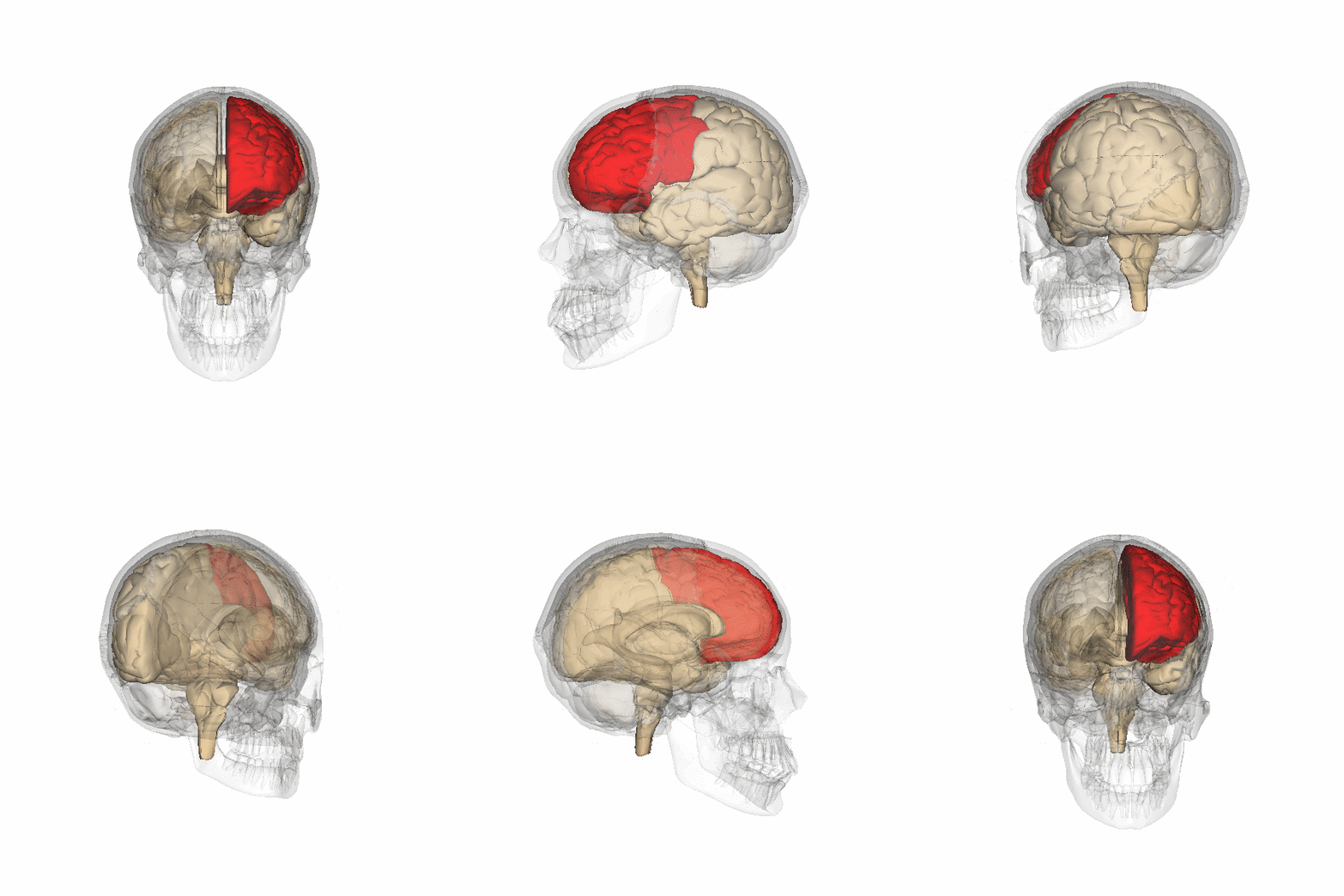
Book definition: “The region at the front of the cerebral cortex that includes the motor cortex and the prefrontal cortex. (p. 15)”. Separated from the parietal lobe by the central sulcus and from the temporal lobe by the lateral sulcus, the frontal lobe plays a key role in the ability to project consequences from actions, choose between “good” and “bad” (i.e. conscience) and override or suppress socially unacceptable responses. It is not fully developed until the late 20s, which in part explains the behavior of children and teenagers. Also contains the primary motor cortex, which regulates bodily movement, as well as most of the dopamine-sensitive neurons in the cortex, which are associated with reward, attention, short-term memory, planning and motivation.
Electroencephalography (EEG)
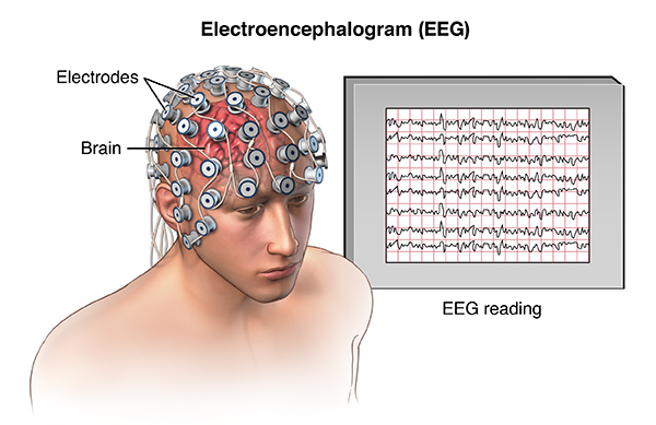
Book definition: “Measurement of electrical activity of the brain, measured by electrodes on the scalp. (p. 20)” This technique has limited spatial resolution due to skull and brain tissue distorting the electrical field, but high temporal resolution and ability to provide a continuous measure of activity in the millisecond range.
Introspection
Book definition: “A methodology much practiced at the turn of the 20th century in Germany that attempted to analyze thought into its components through self-analysis. (p. 4)” Commonly attributed to the German philosopher Wilhelm Wundt, introspection is, in essence, the examination of one’s own conscious thoughts and feelings.

Basal ganglia
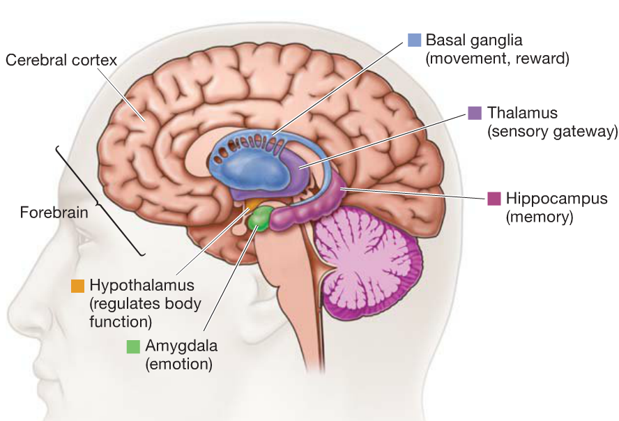
Book definition: “Subcortical structures that play a critical role in the control of motor movement and complex cognition. (p. 16)” The greatest source of insight into the functions of the basal ganglia come from the studying of two neurological disorders that affect the area - Parkinson’s disease and Huntington’s disease - both of which involve impaired motor control. In addition, the basal ganglia have been linked to both eye movement and motivation, where things such as food, sex and drugs elicit a response in a local dopamine system. Damage to this particular subarea can produce a state of apathy.
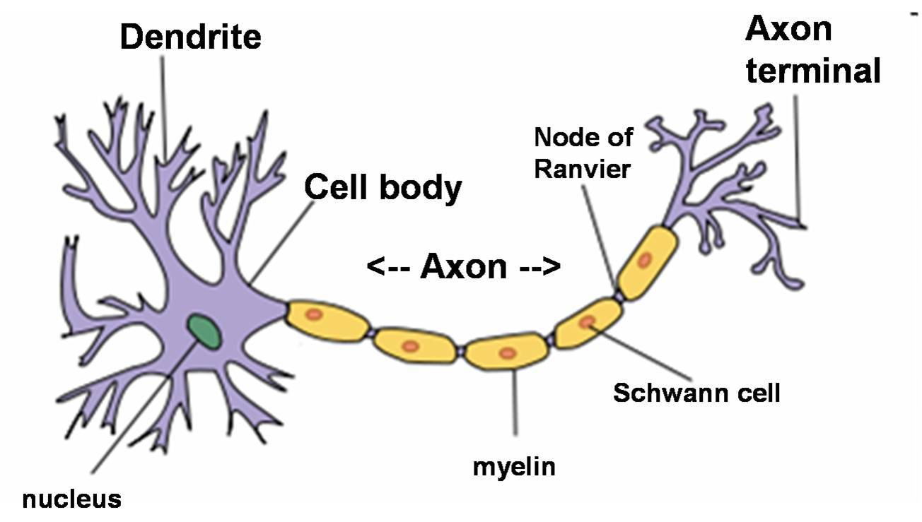
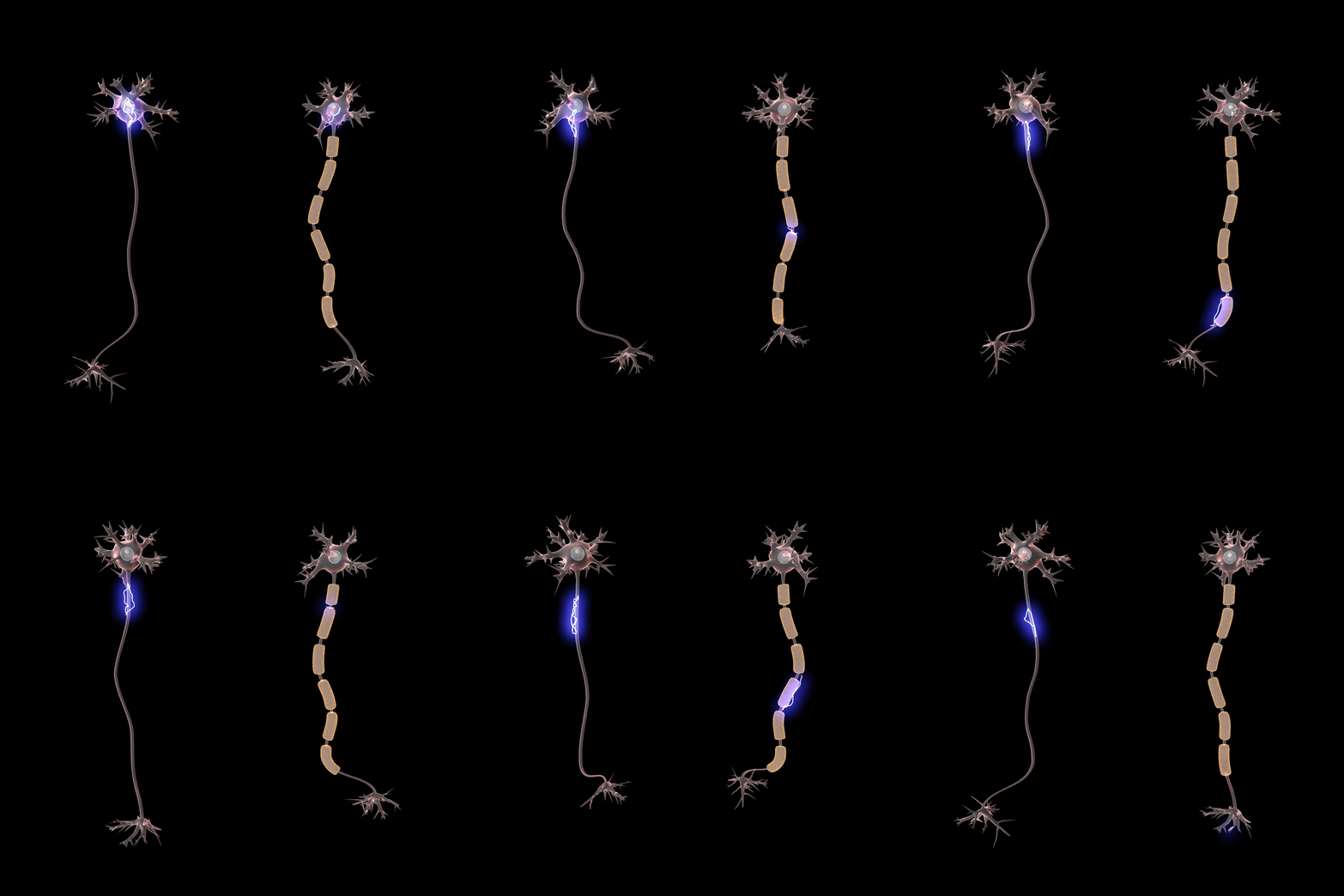
Axon
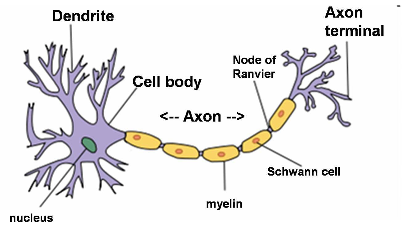
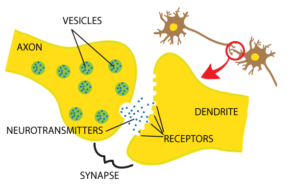
Book definition: “The part of a neuron that carries information from one region of the brain to another. (p. 12)” Axons connect, or synapse, to the tree-like dendrites of other neurons. Axons can be myelinated, meaning they become surrounded by a layer of fat that functions as electrical insulation, making the electrical impulses passing through them faster.
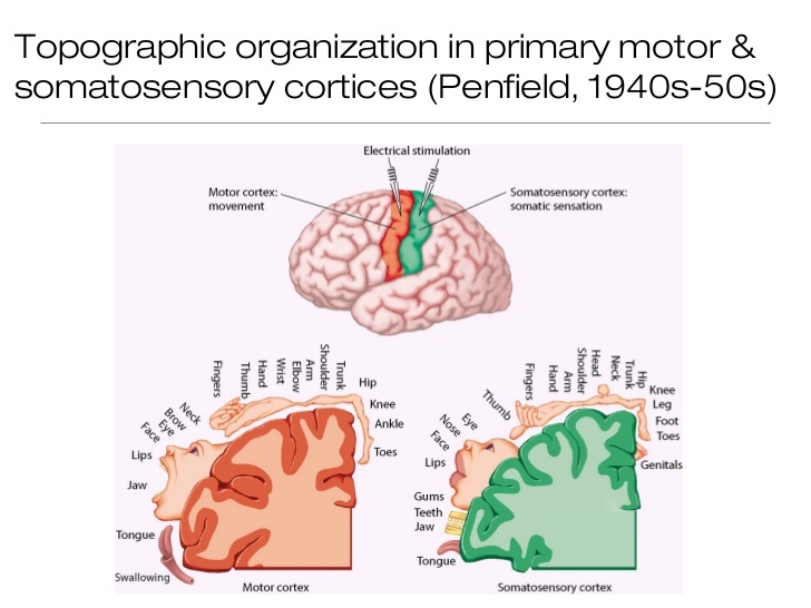
Cognitive neuroscience
Book definition: “The study of the neural basis of cognition. (p. 10)” Cognitive neuroscience was coined by George Miller and Michael Gazzaniga toward the end of the 1970’s. It emerged as an interdisciplinary study of mainly neuroscience, psychology and computer science, with a focus on the scientific study of the biological processes and neuronal connections that underlie cognition and mental processes.
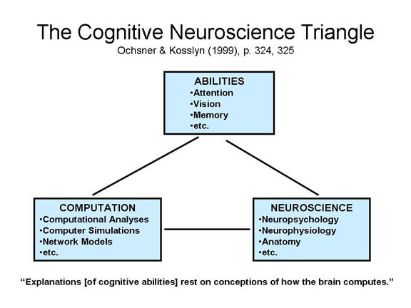
Aphasia
Book definition: “An impairment of speech that results from a brain injury. (p. 17)” Examples: Fluent aphasia (also known as Wernicke’s aphasia), is caused by damage to Wernicke’s area and results in problems understanding and producing meaning (semantics) in written and spoken language, but with an otherwise intact grammar and prosody (syntax). Non-fluent aphasia (also known as Broca’s aphasia), is caused by damage to Broca’s area and results in problems creating fluent and syntactically correct sentences (so-called ‘telegraphic speech’).
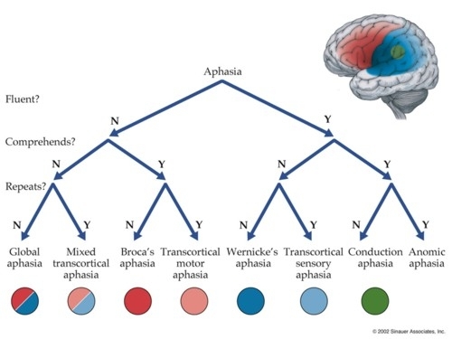
Topographic organization
Book definition: “A principle of neural organization in which adjacent areas of the cortex process information from adjacent parts of the sensory field. (p. 18)”
Excitatory synapse
Book definition: “A synapse in which the neurotransmitters decrease the potential difference across the membrane of the neuron. (p. 12)”
Blood oxygen level dependent (BOLD) response
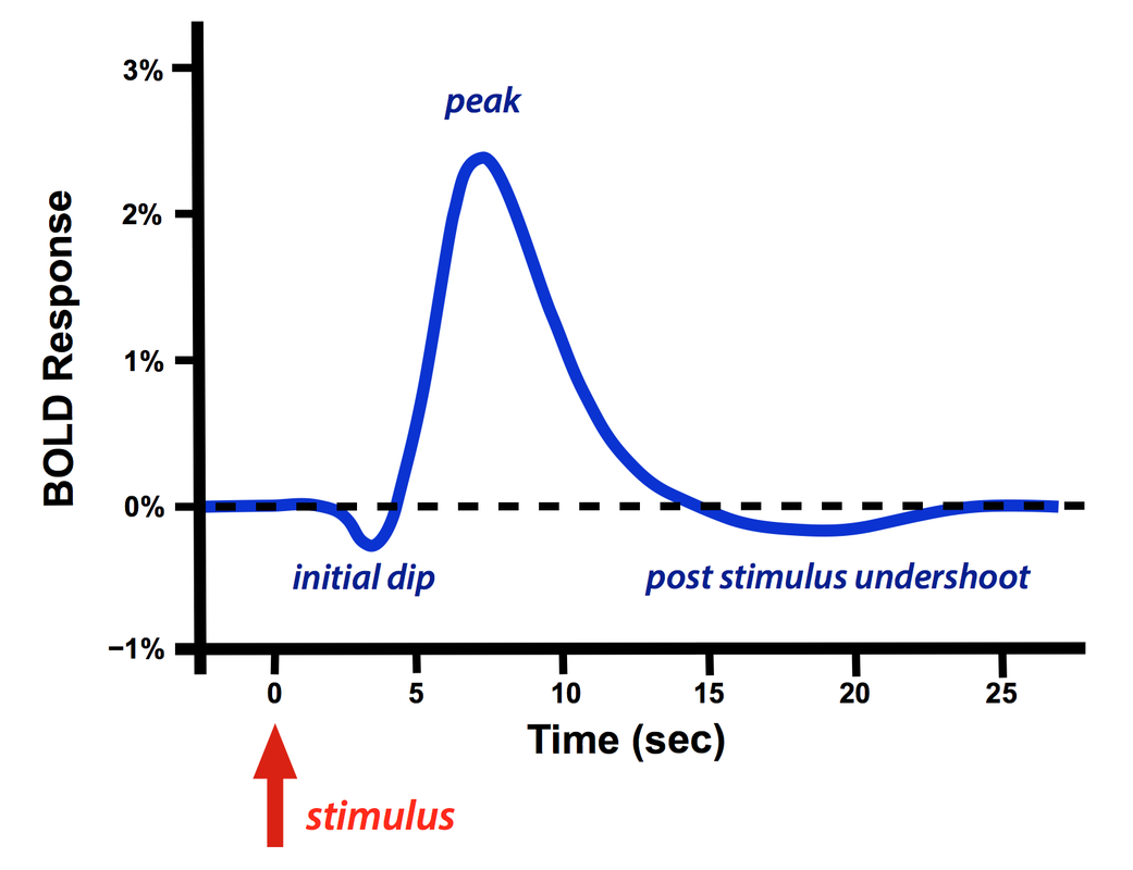
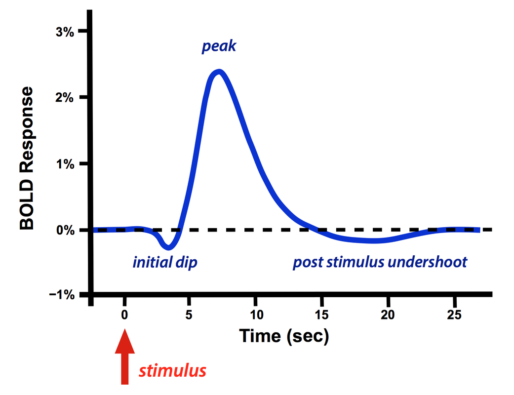
Book definition: “A measure obtained in fMRI studies of the amount of oxygen in the blood. (p. 24)” As neurons consume oxygen, they convert oxyhaemoglobin to deoxyhaemoglobin, which in turn distorts the magnetic field of the brain. By measuring this distortion, the amount of oxygen in the blood (and thereby the activity of the neurons) can be inferred.
Haemodynamic response
Book definition: “The increased flow of oxygenated blood to a region of the brain that has greater activity – the basis of fMRIbrain imaging. (p. 21)” As neurons consume oxygen, they convert oxyhaemoglobin to deoxyhaemoglobin, which in turn distorts the magnetic field of the brain. By measuring this distortion using fMRI, the amount of oxygen in the blood (and thereby the activity-level of the neurons) can be inferred.
Functional magnetic resonance imaging (fMRI)
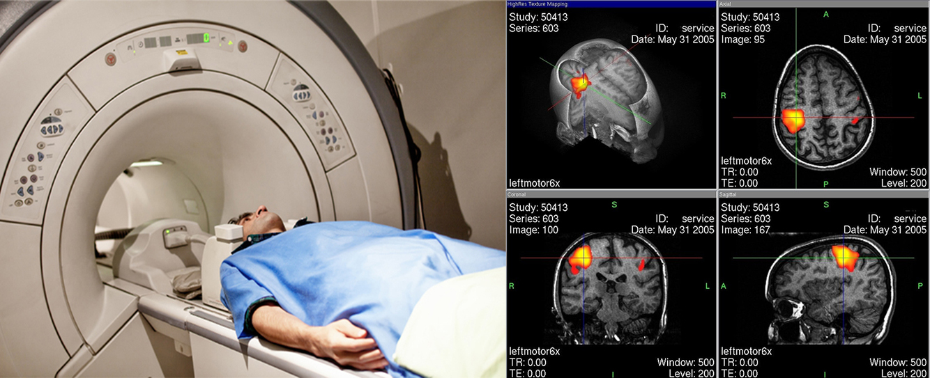
Book definition: “A method for determining metabolic activity by measuring the magnetic field produced by the iron in oxygenated blood. (p. 21)” When neurons consume oxygen, oxyhaemoglobin is converted to deoxyhaemoglobin, which produces distortions in the magnetic field. By assessing this distortion, the concentration of oxygen in the brain (also called the BOLD-response) can be measured. High spatial resolution at about 1 millimeter and medium temporal resolution, detecting changes after about 2-3 seconds. Limitations include patients feeling claustrophobic or being disturbed by the noisy MRI-scanner, while small movements (like answering a question) can distort the BOLD-signal.
Synapse
Book definition: “The location at which the axon of one neuron almost makes contact with the dendrite of another neuron. (p. 11)” The act of creating the connection between the dendrite and the axon is also called “synapsing”.
Empiricism
Book definition: “The position that all knowledge comes from experience in the world. Compare nativism. (p. 4)” Also known as the ‘blank slate view’. This is in contrast to the opposing nativism view, which suggests that humans are born with certain knowledge about language already encoded in our genes.

Myelin
Book definition: “ – ” An insulating layer of fat around some axons (called ‘myelinated axons’) which functions as electrical insulation, making the electrical signal – or action potential – move faster through the axon. Myelination is essential to the proper functioning of the nervous system, and the loss of this layer through demyelination is the hallmark of several neurodegenerative autoimmune diseases such as multiple sclerosis.
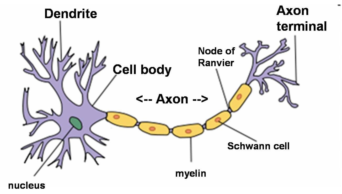
Cell body (also called soma)
Book definition: “ – “ The spherical center of a neuron in which the neuron’s nucleus lies. This connects to dendrites, which bring information to the neuron, and to the axon, which in turn sends information to other neurons by connecting, or synapsing, to their dendrites.
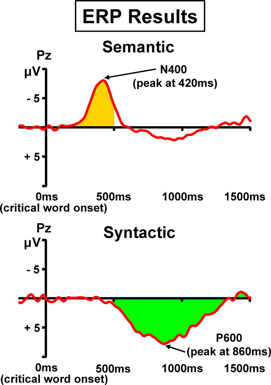
Event-related potential (ERP)
Book definition: “Measurement of changes in electrical activity at the scalp in response to an external event. (p. 21)” This brain imaging technique uses several scalp electrodes, like in EEG, to record the pattern of electrical brain activity during a repeated presentation of the same stimulus. This produces a single waveform, which allows working out the timing of various cognitive processes. Limited spatial resolution due to skull and brain tissue distorting the electrical field, but high temporal resolution and ability to provide a continuous measure of activity in the millisecond range.
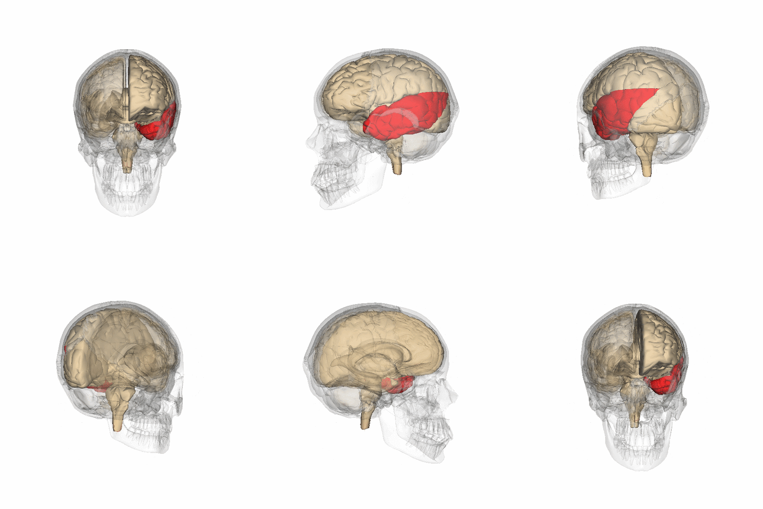
Temporal lobe
Book definition: “The region at the side of the cerebral cortex that contains the primary auditory areas and controls the recognition of objects. (p. 15)” Separated from the frontal lobe by the lateral sulcus, the temporal lobe holds the primary audio cortex (involved in auditory perception and semantics-processing) the hippocampus (involved in memory storage) and Wernicke’s area (involved in the comprehension of written and spoken language). It also shares the fusiform gyrus (involved in facial recognition) with the frontal lobe.
Split-brain patient
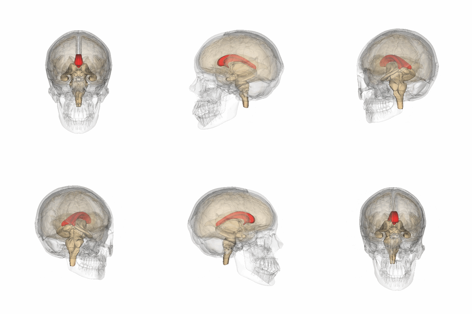
Book definition: “A patient who has had surgery to sever the corpus callosum, which connects left and right hemispheres. (p. 17)” The procedure, known as a “corpus callosotomy,” severs the connection between the left and right brain hemispheres. This is used as a last resort to alleviate powerful epileptic seizures caused by sporadic electrical storms in the brain.
Prefrontal cortex
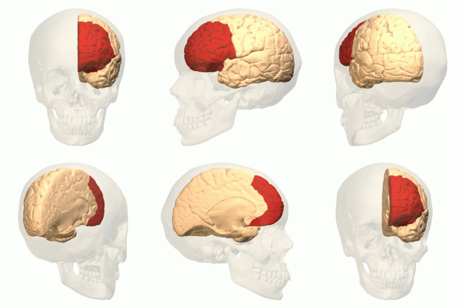
Book definition: “The region at the front of the frontal cortex that controls planning and other higher level cognition. (p. 15)” The prefrontal cortex is heavily involved with top-down processing – i.e. behavior guided by internal states or intentions, such as with personality expression, decision making and moderating social behavior. Functions carried out by the prefrontal cortex are called executive functions and include the abilities to differentiate among conflicting thoughts, considering consequence from current actions, predicting outcomes and suppressing socially unacceptable urges.
Artificial intelligence (AI)
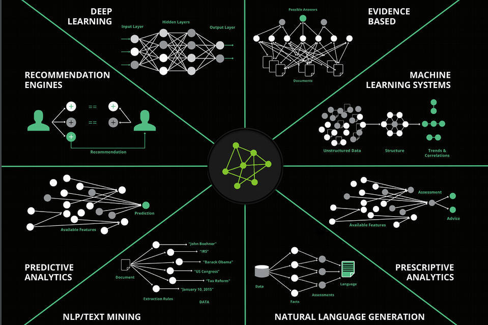
Book definition: “A field of computer science that attempts to develop programs that will enable machines to display intelligent behavior. (p. 1)”
Rate of firing (ROF)
Book definition: “The number of action potentials, or nerve impulses, an axon transmits per second. (p. 13)”
Broca’s area
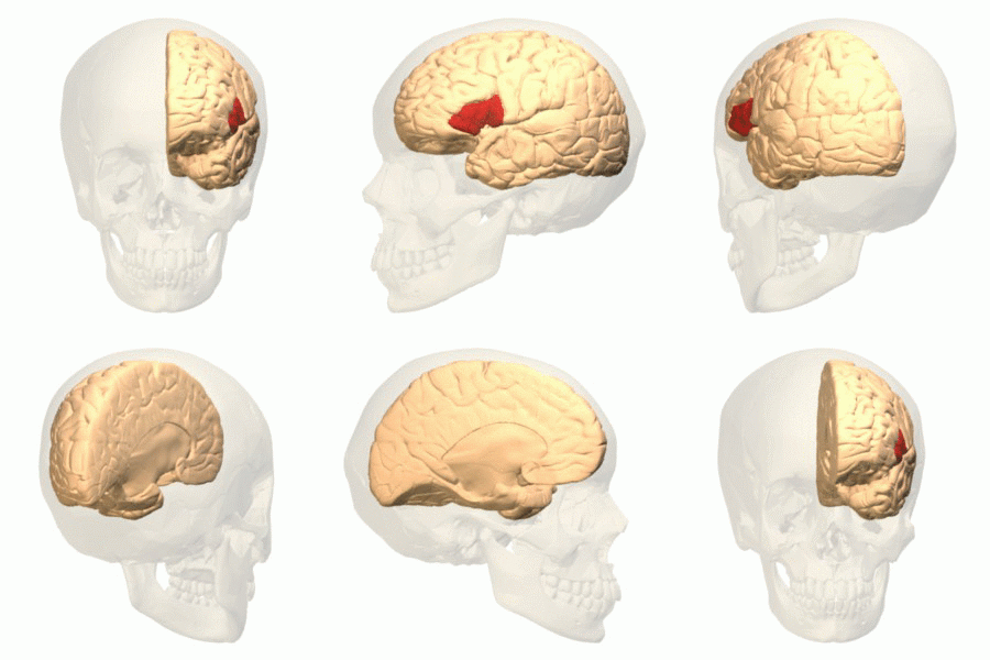
Book definition: “A region in the left frontal cortex that is important for processing language, particularly syntaxin speech. (p. 17)” Damage to this area can lead to non-fluent aphasia, which results in an inability to create fluent phrases out of otherwise meaningful words (so-called ‘telegraphic speech’) – e.g. this actual patient quote: “The woman… wants to… The girl wants to… the flowers and the woman.” See also Wernicke’s area and fluent aphasia.
Dendrite
Book definition: “The branching part of a neuron that receives synapses from the axons of other neurons. (p. 11)” Due to the tree-like shape of dendrites, their branching is called ‘arborization’ (‘arbor’ being Latin for ‘tree’). The connections made between axons and dendrites are called synapses.