Analyzing Cells
No description yet. This curated deck is ready for daily study.
Card preview (24)
Transmitted Light
Bright-field / phase contrast / differential interference contrast -DIC Passed through sample
Fluorescence
Specific localization of organelles, macromolecular structures, and molecules within cells. add tag to protine
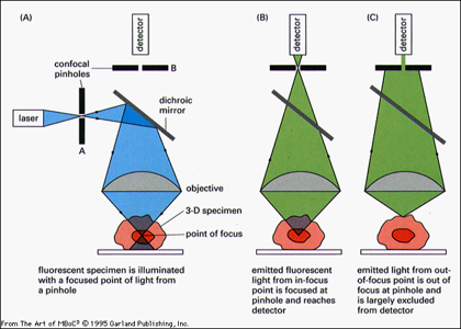
Laser Scanning Confocal Microscopy
Similar to Bright-field and Fluorescence, but increased clarity due to removal of out-of-focus light. pinpoint illumination
Limit of resolution
Limiting separation at which two objects appear distinct. Depends on • wavelength of light • Numerical Aperture of lens system
Numerical Aperture of lens system
higher NA better resolution (NA affects light gathering ability, related to angle of cone of light (greater with greater angle) that can enter it and refractive index of medium (how light bends/scatters). Refractive index is the ratio of speed of light in vacuum to speed of light in a particular medium
Why oil immersion lenses give betteer resoluton
air glass oil 1.00 1.51 1.52 because glass and oil have the same refractive index, no interference - less light scattering - more light passing through objective lense
top vs bottom lense
top - objective lense bottom - condensor lense
Bright Field
: Light transmitted straight through specimen. View living cell, but poor contrast.
Phase of light passing through a living cell is changed according to cell’s
refractive index
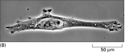
Phase contrast
: Phase alterations of light transmitted through the specimen are translated into brightness changes. neg. - halow effect extra brightness - obstructs view
DIC
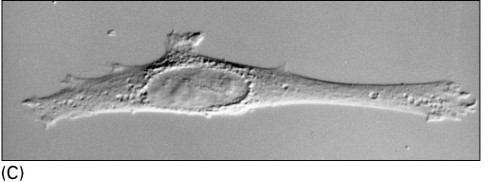
(Differential Interference Contrast): Highlights edges where there is a steep change in refractive index. neg. - shadow effect
H&E stain
acidic and basic properties
chemical stain
specificaly bind to specific structure neg- introduce artifacts, no longer living
How tissue is prepaired
Fixative applied (preserve sample) Embedded in paraffin wax or resin (hardened to form a solid block) Cut into thin slices by a microtome (~.5 um for light microscopy)
Fluorescent probes
real time without killing sample folrofloresence- tag - can be exctived with obsoption of light - excited state - unstable - beack to ground (emits light - larger wavelength than emited)
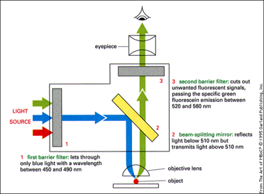
Fluorescent microscope
beam splicer - make sure splited light light on one side not transmited - reglexted and twards
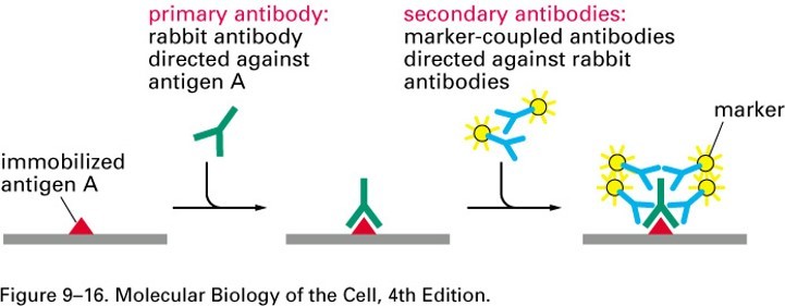
Antibodies labeled with fluorescent dye
Used to detect specific proteins or other molecules in cells and tissues need second antibody - ampldy singnl - brighter
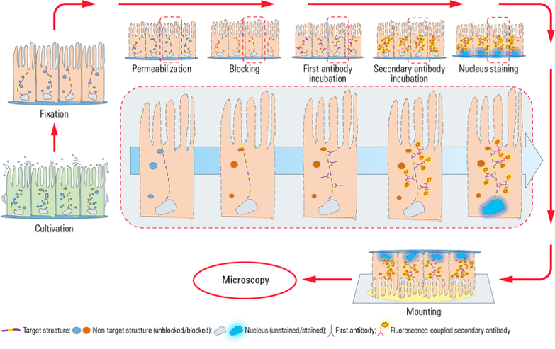
STEPS: INDIRECT IMMUNOFLUORESCENCE
• Fixation: Preserve and kill cells. Maintain cellular morphology (chemical crosslinkers: formaldehyde) • Permeabilization: Enable antibodies to cross cell membrane (detergent disrupts cell membrane) • Blocking: minimize unspecific binding of the primary antibody within the cell (normal serum, proteins such as casein) • Incubate with primary Antibody: Primary antibody binds to protein of interest • Incubate with secondary Antibody (fluorescent label): Binds to primary antibody (signal amplified) • Stain: localization within cell • Mount: fix sample on slide (prevent dehydration)
Green fluorescent protein (GFP),
isolated from jellyfish. Used to monitor gene expression. GFP joined to fly promoter: active only in specialized set of neurons view living processes - genetic engenerign - endogenous production
Superresolution Fluorescence
• Florsent - bulurry - differnt debths Overcome limit of resolution (light microscopy) to 20 nm, order of magnitude improvement! If specimen contains large number of adjacent fluorescent molecules, blurry image. supress - turn on and off florsences
PALM
Photoactivated localization microscopy: supress - turn on and off florsences
TIRF:
Total internal reflection fluorescence
Fluorescence Microscopy Pros and Cons
Pros: Label specific proteins within cell using fluorescently labeled antibodies, label multiple molecules to observeerve interaction and localization. Super resolution fluorescence microscopy-limit of resolution is 20nm (order of magnitude below classic diffraction limit to resolution) Cons: Fluorescence is not permanent. Photobleaching occurs as samples are viewed
Confocal Microscopy
excluding out-of-focus light - illuminating part - at specidic depth used to resolve 3D structures of numerous objects, including networks of cytoskeletal fibers in cytoplasm and arrangement of chromosomes and genes in the nucleus. uses pinhile - only light from focoused area gets through and lazer also goes through pinhole - specific point on sample