Biology Quiz: Microscopes & Parts of a Cell
No description yet. This curated deck is ready for daily study.
Card preview (24)
What do microscopes do?
-Main benefit is resolution, the ability to see fine detail
1595 Zaccharias and Hans Janssen (Holland)
• Produced crude microscope • Used a 2-lens system
1609 Galileo (Italy)
-Built crude compound microscope
1600’s Anton Van Leeuwenhoek (Holland)
• Built simple single lens microscope • 1st to see unicellular motion
1665 Hooke (England)
• built compound microscope • First person to use the word “cell” (cork) • 3 lens system
1930 Hillier & Prebus (Canada)
-Built 1st electron microscope
1940’s
-first scanning electron microscope
Compound research microscope (common)
• Image produced by light through specimen • 1 eyepiece/ocular (0-4 objective lenses) • Transparent specimens used • 1000X
Dissecting Microscope [compound microscope]
• 2 oculars/eyepieces • 2 light sources • To look at solid objects • 85X
Pros & Cons of Compound Microscopes
Pros: -images produced in color, 3D, living materials can be used Cons: • low magnifacations • poor resolution at high magnifacations
Parts of a microscope
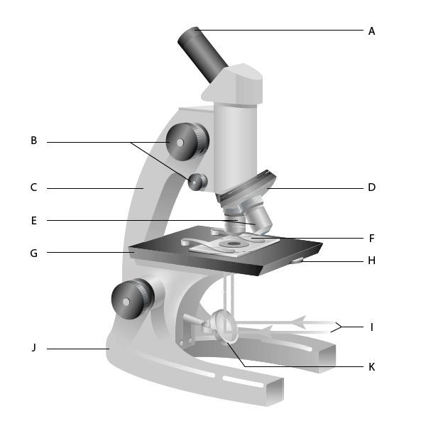
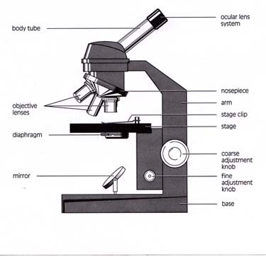
Transmitted light
Light passes through specimen
Incident Light
Light reflects off specimen
Parfocal
When microscope is focused on one power, it should still be in focus when swtiched
Inverted Image
Upsidedown and backwards (‘e’)
Virtual Image
Image is the same as it is
Field of View
• Diameter of what you see • Measured in microns (μ) • Descreases as magnifacation increases • Forumla: known f.o.v/(unknown mag/ known mag)* *known mag= image ocular x objective for each*
mm to microns
1mm x 1000 = 1 micron
Actual Size
= field diameter/fit number
Calculate the Scale
scale = actual size/ drawing
Magnifacation
1.) number of times microscope enlarges specimen magnifacation= drawing/actual sizw
Electron Microscope
• Uses beam of electrons not lightwaves • No colour • Able to see great detail
Transmission Electron Microscope (TEM)
-Beam of electrons pass through stained tissue imbedded in plastic Pros: • very high mag. • high resolution (internal detail seen) Cons: • 2D • B&W
Scanning Electron Microscope (SEM)
• scans surface of specimen • image produced by electrons reflected off surface onto screen • Often coat specimen gold for sharp images Pros: • High magnifacation • 3D B&W Cons: -specimen has to be dead